Case Report
Austin J Clin Case Rep. 2023; 10(1): 1274.
An Unusual Bilateral Cicatrizing Conjunctivitis in Severe Ocular Rosacea: Case Report
Elbelidi H*, Saoiabi Y and Cherkaoui L
Department of Ophthalmology, Hopitaldes Spécialités, Rabat, Morocco
*Corresponding author: Elbelidi HDepartment of Ophthalmology, Hopitaldes Spécialités, Rabat, Morocco
Received: January 31, 2023; Accepted: March 06, 2023; Published: March 13, 2023
Abstract
Rosacea is a chronic progressive disease of unknown cause that affects the eyes and facial skin. Ocular rosacea is often missed when ophthalmologists do not adequately examine a patient’s face during eye exams. If treatment is delayed due to undiagnosed rosacea, serious eye complications and blindness can occur.
We present a case of 35-year-old female, followed up for rosacea and severe depressive disorder, presented with bilateral, symmetrical blinding cicatricial conjunctivitis after neglected ocular self-injury.
Keywords: Rosacea; Cicatrizing; Conjunctivitis
Introduction
Rosacea is a multifactorial, chronic, inflammatory dermatological condition that affects forehead, eyelids, cheeks, nose and chin [1]. In most cases, the disease progresses in a relapsing and course, with exacerbations that can be triggered by exposure to ultra-violet radiation, spicy foods, or heat [2]. It is often overlooked by ophthalmologists, as ocular manifestations precede cutaneous disease in 15% of cases [3]. More than 50% of individuals have ocular abnormalities, which can range from minor dryness and irritation, blepharitis and conjunctivitis to sight-threatening keratitis [4].
Ocular rosacea has a significant psychosocial impact and can be potentially blinding, as a result, recognizing the condition is an important part of management [5,6].
Here, we report a patient with ocular rosacea, who had severe sight-threatening bilateral cicatrizing conjunctivitis.
Case Report
A 35-year-old woman presented with a five-month history of irritation, blepharospasm and redness in both eyes. She was diagnosed with severe depression two years ago, with the notion of two suicide attempts, self-harm and neglected eye self-injury. She denied any other autoimmune disease, skin disorder, drug allergy, eye procedure.
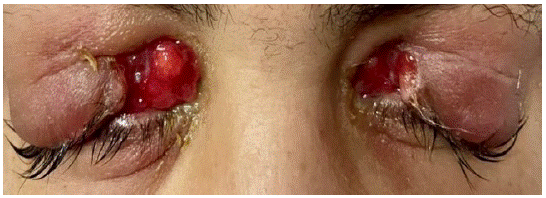
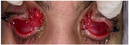
The visual acuity was light perception in both eyes. The External examination revealed in both eyes inflamed upper eyelids, notably there was a solution of continuity of the internal cantus, and through this we could see a very inflamed conjunctiva with rounded granular lesions and secretions (Figure 1 & 2). Slit lamp examination showed a conjunctival injection, foreshortening of the conjunctival fornices was present along with 360 degrees of continuous symblepharon in both eyes. A clinical diagnosis of chronic cicatricial conjunctivitis was made.

Figure 1: Photograph of both eyes showing cuts in the medial cantus.

Figure 2: Photograph of both eyes showing a very inflamed conjunctiva with granuloma.
To obtain more definitive diagnosis, conjunctival biopsy was performed for granular lesions and conjunctival adhesions. Histopathologic examination of the specimen revealed an ulcerated squamous and columnar epithelium, covered with fibrin-leukocyte masses. The chorion showed neovascular proliferation perpendicular to the surface with a pleomorphic inflammatory response (mixed with a granulomatous giant cell component) in contact with the foreign body.
Following the biopsy results, dexamethasone 0, 1% + tobramycin 0, 3% drops were begun 4 times daily. Comprehensive investigations, including complete blood count, erythrocyte sedimentation rate, c-reactive protein, quantiFERON-TB Gold, serum angiotensin-converting-enzyme, serum lysozyme level, serum calcium level, antinuclear antibodies, anti SSA/Ro antibodies, anti-La/SSB antibodies, anti-Smith antibodies, anti-Jo 1antibodies, CCP antibodies, rheumatoid factor blood test and Anti-neutrophil cytoplasmic antibodies were normal. Computed tomography of the chest was without any particularity. The diagnosis of post-ocular trauma cicatrizing conjunctivitis in a patient treated for rosace was maintained.
The patient was treated with doxycycline 100 mg capsule once daily combined with dexamethasone 0, 1% eye drops twice a day, cyclosporine 0.5% eye drops 4 times a day and preservative free lubricants six times a day, in both eyes.
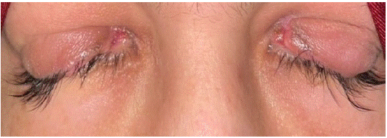
After two months of treatment, there was a clear improvement in the inflammatory signs, with regression of the palpebral oedema, hyperthermia and conjunctival granulomas, as well as healing of the internal cantus lesions (Figures 3 & 4).

Figure 3: Photograph of both eyes showing a clear regression of inflammatory signs after 2 months of treatment.

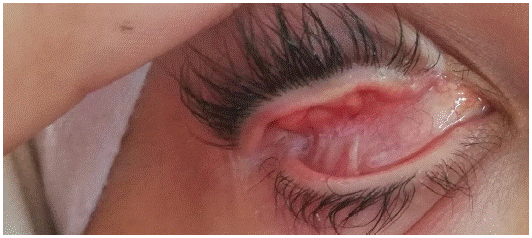
Figure 4: Photograph of the right eye showing symblepharons.
After two months of treatment, the inflammatory symptoms improved significantly, and the patient was scheduled for symblepharon release and fornix reconstruction.
Discussion
Ocular rosacea affects up to 58% of patients with cutaneous rosacea [1]. However, the extent of cutaneous involvement is not necessarily directly related to the severity of ocular involvement [7]. In patients with diagnostic features, a diagnosis of ocular rosacea should be considered independently of the cutaneous disease. Bilateral ocular manifestations are common, but unilateral or sequential changes can occur [3].
The most common manifestations are eyelid margin telangiectasias, meibomian gland dysfunction, and posterior blepharitis [8]. The cornea is involved in approximately one-third of ocular rosacea cases [3]. Episcleritis, scleritis and anterior uveitis are also reported [8]. The chronic inflammation associated with ocular rosacea may result in chronic nonspecific conjunctivitis, primarily affecting the interpalpebral region [9]. Conjunctival scarring, phlyctenulosis, and conjunctival granulomas may occur [3]. Sterile corneal inflammation, sclerokeratitis and scarring conjunctivitis are the main causes of vision loss in this disease [10].
Our case concerns a patient followed up for rosacea and severe depressive disorder, presented bilateral and symmetrical blinding cicatricial conjunctivitis. To our knowledge, this is the first reported case of bilateral cicatricial conjunctivitis in the context of ocular rosacea.
In rosacea, ocular involvement is less easily recognized and often remains underdiagnosed despite serious complications [3]. Ocular changes may precede skin changes in approximately 20 percent of patients; however, skin lesions appear first in half of patients, and both manifestations occur in one-third of patients [4]. There are no laboratories or histopathological tests to confirm the diagnosis of ocular rosacea. Diagnosis depends on evaluation of clinical symptoms. Response to a trial of oral tetracycline may help confirm the initial diagnosis [4]. If treatment is delayed due to undiagnosed ocular rosacea, serious ocular complications and permanent vision loss may occur [11].
The differential diagnosis of cicatricial conjunctivitis includes autoimmune disorders such as systemic lupus erythematosus, post-infection, trauma, thermal and chemical burns, or iatrogenic (Stevens-Johnson syndrome). Several underlying causes can result in significant systemic morbidity and even mortality. Therefore, adequate and timely diagnosis of the underlying disease is crucial and can save lives and allow for appropriate treatment [12].
The management of cicatricial conjunctivitis is according to the underlying etiology. However, it may not be always possible to identify the underlying cause and in case our case, we should manage contributing factors such as ocular self-injury, dry eye, rosacea disease.
Conclusion
Ocular rosacea should be kept in mind in the differential diagnosis of chronic cicatrizing conjunctivitis. It can be a diagnostic challenge in the case of lack of skin lesions. Early diagnostic and treatment will provide symptomatic relief and reduce the risk of complications.
Declaration of Conflicting Interests
The author(s) declared no potential conflicts of interest with respect to the research, authorship, and/or publication of this article.
Statement of Consent
The authors have received consent from the patient to publish the attached images and case details.
References
- Starr PA, Macdonald A. Oculocutaneous aspects of rosacea. Proc R Soc Med. 1969; 62: 9-11.
- Two AM, Wu W, Gallo RL, Hata TR. Continuing medical education. Rosacea Part I. Introduction, categorization, histology, pathogenesis, and risk factors. J Am Acad Dermatol. 2015; 72: 749-758.
- Akpek EK, Merchant A, Pinar V, Foster CS. Ocular rosacea: patient characteristics and follow-up. Ophthalmology. 1997; 104: 1863-1867.
- Browning DJ, Proia AD. Ocular rosacea. Surv Ophthalmol. 1986; 31: 145-58.
- Incel Uysal P, Akdogan N, Hayran Y, Oktem A, Yalcin B. Rosacea associated with increased risk of generalized anxiety disorder: a case – control study of prevalence and risk of anxiety in patients with rosacea. An Bras Dermatol. 2019; 94: 704-709.
- Oussedik E, Bourcier M, Tan J. Psychosocial burden and other impacts of rosacea on patients’ quality of life. Dermatol Clin. 2018; 36: 103-113.
- Crawford GH, Pelle MT, James WD. Rosacea: I. Etiology, pathogenesis, and subtype classification. J Am Acad Dermatol. 2004; 51: 327-341.
- Ghanem VC, Mehra N, Wong S, Mannis MJ. The prevalence of ocular signs in acne rosacea: comparing patients from oph- thalmology and dermatology clinics. Cornea. 2003; 22: 230-233.
- Vieira AC, Hofling-Lima AL, Mannis MJ. Ocular rosacea—a review. Arq Bras Oftalmol. 2012; 75: 363-369.
- Erzurum SA, Feder RS, Greenwald MJ. Acne rosacea with keratitis in child- hood. Arch Ophthalmol 1993; 111: 228-230.
- Celiker H, Toker E, Ergun T, Cinel L. An unusual presentation of ocular rosacea. Arq Bras Oftalmol. 2017; 80: 396-398.
- Vazirani J, Donthineni PR, Goel S, Sane SS, Mahuvakar S, et al. Chronic cicatrizing conjunctivitis: A review of the differential diagnosis and an algorithmic approach to management. Indian J Ophthalmol. 2020; 68: 2349-55.