Case Report
Austin J Clin Case Rep. 2023; 10(3): 1283.
Angiomyxoma – A Rare Finding Associated with Congenital Cyst Malformation of the Umbilical Cord
Yuvaraj H; Aboda A*; Pettigrew I; McCully B
Department of Obstetrics & Gynaecology, Mildura Base, Public Hospital, Australia
*Corresponding author: Ayman Aboda Department of Obstetrics & Gynaecology, Mildura Base, Public Hospital, Australia. Email: aymanaboda@hotmail.com
Received: April 21, 2023 Accepted: May 15, 2023 Published: May 22, 2023
Abstract
Although rare, umbilical cord cysts may be associated with prenatal aneuploidy and foetal developmental anomaly. If unresolved, they may affect growth support with a risk of progressive or sudden vascular compromise. In this report, we discuss the case of a 33-year-old woman who presented with a large septate umbilical cord cyst detected incidentally during routine morphology scanning. The patient had low-risk first-trimester combined screening, later confirmed by Non-Invasive Prenatal Testing (NIPT). She went on to have detailed antenatal ultrasound surveillance and delivered successfully at term. The case highlights the importance of multidisciplinary collaboration with tertiary partners to align clinical care with best practices and evidence-based decision-making. This is particularly relevant in regional settings where the workforce may be strained or transient and the knowledge base of rare conditions less robust.
Keywords: Angiomyxoma; Umbilical cord cysts; Congenital abnormality; Aneuploidy
Case Presentation
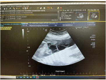
A 33-year-old Caucasian woman with a history of three uneventful deliveries attended a regional hospital for antenatal care. Her 12-week ultrasound scan showed a normal intrauterine pregnancy consistent with gestational age. The nuchal translucency was within normal limits, and no adnexal cysts or masses were detected. Non-Invasive Prenatal Testing (NIPT) results confirmed low risk for trisomy 13, 18, and 21. Routine antenatal screening, including viral serology, was normal. At 20 weeks, she had routine second-trimester scan which showed normal fetal morphology, an anterior, non-praevia placenta with normal cord insertion. Unexpectedly, a large cystic septate structure measuring 65x32mm was observed within the umbilical cord. The findings were reviewed by a tertiary scan, confirming two large, avascular umbilical cysts that measured 62.4x40.6x41.2mm and 53.3x48.3x51.6mm. There was no evidence of cord compression or impaired vascular flow. The diagnosis was consistent with two septate umbilical cord cysts, likely arising as areas of oedema within the Wharton jelly (Figure 1).

Figure 1: Umbilical cord cysts at morphology USS.
Collaborative discussion with the maternal-fetal medicine team advocated a plan for ongoing care with the regional obstetric service, ensuring four-weekly USS surveillance to monitor fetal growth and well-being and to assess any change in size or evidence of cord vessel compression. The patient went on to have an otherwise uneventful antenatal journey. The cysts remained but were uncomplicated. The foetus grew well, exceeding expected growth velocities (Figure 2).

Figure 2: Umbilical cord cyst at 36 weeks.
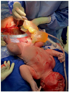
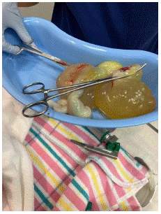
At 41 weeks and four days, the patient presented in spontaneous labour. She had an uneventful first stage, monitored with continuous CTG tracing. The foetal heart was normal throughout. The second stage was prolonged. The presenting part was high and in an occipital-posterior position which was not suitable for assisted vaginal delivery. She had an emergency Caesarean section under spinal anaesthesia. The baby was delivered in good condition. The umbilical cord was inspected and milked towards the baby to ensure no abdominal contents herniated within the cysts prior to clamping. The baby did well, with APGAR scores of nine at 1 minute and ten at 5 minutes, and the birth weight was 4.71kg. Cord blood lactate was normal. The placenta weighed 595g. The umbilical cord measured 230mm with a diameter of 14mm to 28mm (Figures 3-6).

Figure 3:

Figure 4:

Figure 5: Overall survival, autologous stem cell transplant (ASCT) versus no ASCT (p=0.12).

Figure 6:
The cord was fixed in formalin and sent for histopathology. The findings confirmed three vessels, with a proliferation of smaller vessels arising from one of the umbilical arteries. An angiomyxoma was identified though it was uncertain whether this was a reactive change or a separately arising neoplasia. Wharton's jelly was markedly oedematous and contained several smooth-walled cystic structures ranging from 8mm to 110x83x53mm. All contained clear, viscous fluid with no nodules or other lesions. There was no evidence of other significant pathology in the placenta or membranes.
Discussion
Umbilical cord cysts are rare anomalies found during pregnancy. They are identified in approximately 2-3% of first-trimester ultrasound scans and frequently resolve by the second trimester [1,2]. They are classified as either true or pseudocystic. The former arise from embryonic remnants, such as the allantois or omphalomesenteric duct, and have a characteristic epithelial lining. They are associated with an increased risk of fetal anomalies, spontaneous abortions, and aneuploidies (chromosomal abnormalities) such as trisomy 13 and 18 [3,4]. When arising from the allantois, true cysts may be associated with urachal anomalies that may connect to the fetal bladder [5]. They may also be contiguous with the abdominal wall, where they can rapidly expand, causing constriction of umbilical blood flow and foetal compromise [5]. Pseudocysts are more common and present as large, hypoechoic masses near the cord's insertion at the foetal umbilicus. They are often multiloculated and may also occur within free loops of the cord [1]. They do not have an epithelial lining and are formed by localized oedema of Wharton's jelly, the gelatinous substance surrounding and protecting the umbilical cord vessels [3]. Diagnosis of umbilical cord cysts begins with ultrasound imaging; however, differentiation of true or pseudocysts origin may be challenging, with definitive diagnosis only possible following histological examination of the cord after delivery [6]
In general, single cysts possess an excellent prognosis if they resolve in later pregnancy [8]. However, multiple cysts are associated with an increased risk of adverse findings, particularly if they persist beyond 14 weeks [7]. These may include miscarriage, aneuploidies, omphalocele, growth restriction, and VACTERL, which is an acronym for a number of congenital anomalies that may arise synchronously as a result of either genetic or environmental factors [8,9].
Angiomyxoma is a rare, benign soft tissue tumour that arises predominantly in the pelvic and perineal regions. It typically affects women of reproductive age. Although slow-growing and non-metastasizing, the tumour may cause significant morbidity due to its propensity for invasive and proliferative growth. Angiomyxoma arising from umbilical cord vessels is equally rare [10]. Case reports are isolated, so the actual incidence is unknown. They are typically discovered incidentally with no associated clinical significance. Occasionally, however, when large, they may cause compression of adjacent cord vessels leading to impaired foetal vascular support. They may also cause torsion leading to precipitant fetal death [11]. Once discovered or suspected, they are monitored by regular USS and Doppler assessments to ascertain their growth and fetal well-being [11].
Conclusion
Congenital umbilical cord cysts and angiomyxoma are rare conditions that require detailed USS surveillance to monitor complications and ensure ongoing fetal well-being. This report highlights the importance of multidisciplinary collaboration with partnering services, especially in regional areas where the medical workforce may need support to manage rare conditions. By raising awareness, we hope to support the longevity and sustainability of safe patient care in local, known environments by trusted and enabled caregivers.
References
- RE Bohîltea, V Dima, I Ducu, AM Iordache, BM Mihai, et al. Clinically relevant prenatal ultrasound diagnosis of umbilical cord pathology. Diagnostics. 2022; 12: 236.
- EFM Santana, RG Castello, G Rizzo, G Grisolia, Junior EA, et al. Placental and Umbilical Cord Anomalies Diagnosed by Two-and Three-Dimensional Ultrasound. Diagnostics, 2022; 12: 2810.
- EFM Santana, RG Castello, G Rizzo, G Grisolia, Junior EA, et al. Placental and Umbilical Cord Anomalies Diagnosed by Two-and Three-Dimensional Ultrasound. Diagnostics. 2022; 12: 2810.
- BD Rathod, P Tamilarasan. Umbilical Cyst with Edward Syndrome. Indian Journal of Neonatal Medicine and Research. 2016; 4.
- Adesina SB, Bassey K, Bassey EA, Nyong EE, Balogun YA, et al. Giant Cystic Umbilical Cord in a Post-Term Nigerian Neonate. Journal of Diagnosis & Case Reports. 2022; 3: 1-3.
- Kaur N, McKenney AH, Kollikonda S, Karnati S. Changing Course of an Umbilical Cord Mass-Chasing the Diagnosis of Angiomyxoma. Pediatr Dev Pathol. 2022; 25: 558-561.
- LR Campo, RS Cornudella, FG Alderete, Payo CM, Perez PP, et al. Prenatal diagnosis of umbilical cord cyst: clinical significance and prognosis. Taiwan J Obstet Gynecol. 2017; 56: 622-627.
- M Fahmy - Umbilicus and Umbilical Cord. Springer. 2018.
- Q Liu, R Wei, J Lu, H Ding, H Yi, et al. A Retrospective Cohort Analysis of the Genetic Assay Results of Foetuses with Isolated and Nonisolated Umbilical Cord Cyst. Int J Gen Med. 2022; 15: 5775-5784.
- H Göksever, M Celiloglu, A Kupelioglu. Angiomyxoma: a rare tumour of the umbilical cord. J Turk Ger Gynecol Assoc. 2010; 11: 58-60.
- W Sepulveda, LL Simpson. Umbilical cord abnormalities: Prenatal diagnosis and management. Up to Date. 2021.