Orthopedics Case Reports
Austin J Clin Case Rep. 2023; 10(4): 1285.
Functional Outcome of Surgically Managed Acetabular Fractures
Muhammad Inam; Imran Khan*; Asif Khan; Muhammad Shabir; Mian Amjad Ali
Department of Orthopedic and Trauma, Medical Teaching Institute Lady Reading Hospital Peshawar, Pakistan.
*Corresponding author: Imran Khan Department of Orthopedic and Trauma, Medical Teaching Institute Lady Reading Hospital Peshawar, Pakistan. Email: drminamkhan71@gamil.com
Received: May 02, 2023 Accepted: May 27, 2023 Published: June 03, 2023
Abstract
Objective: The objective of study was evaluate the results of open reduction and rigid internal fixation with Reconstruction plates and screws in acetabular fractures.
Material and methods: This prospective case series study was conducted at Orthopedic and Trauma Department of medical Teaching Institute Lady Reading Hospital (LRH) Peshawar Pakistan January 2018 to December 2022 on 21 consecutive patients of either gender with the age range from 18-70 years, having acetabular fracture presenting within one month of injury. Patients with open fracture and trochanteric pin for traction as a treatment modality were excluded from the study. Non-probability consecutive sampling technique was used. Patients were followed for a minimum of 6 months. Clinical grading was done according to D’aubigne and Postel modified by Matta. Pain, gait and range of motion were assessed. Radiological grading was done on last visit according to Matta criteria as excellent good, fair and poor.
Results: There was total 21 patients having age range of 20-60 years with average age of 37.52. Male were 14 (66.7%) while female were 7 (33.3%). The cause of injury was road traffic accident in 13 (61.9%), Fall 6 (28.6%) and Physical Violence in 2 (9.5%) patients. Right side was involved in 13 (61.9%) while left was involved in 8 (38.1%) patients. There was plate loosening in 2 (9.5%) cases. Excellent results were obtained in 11 (52.4%), good in 7 (33.3%), fair in 2 (9.5%) and poor in 1 (4.8%) cases.
Conclusion: Open reduction and internal fixation with 3.5 millimeter reconstruction plate and screws gives Excellent to Good results according to Matta Grading in all fracture of the acetabulam even in osteoporotic bone as well.
Keywords: Acetabulam; Fracture; Internal fixation; Open reduction; Rigid fixation
Introduction
Modernization and industrialization has made more trauma and more road traffic accidents [1]. Trauma can cause multiple injuries to the patients and developed countries has more deaths at the scene of trauma due to high impact [2]. One of the dangerous trauma is the injury to the pelvis which can cause fracture of the acetabulam and if the patient is not resuscitated, s/he may die of blood loss. So management of acetabulam fracture needs priority in trauma patients [3].
Acetabular fracture can occur due to high impact trauma like motor vehicle accident, fall from height or run over injury [4]. Initially such fractures were treated conservatively but with the passage of time and due to complication of conservative management, surgical fixation has been introduced by Judet And Leuternal [5]. They have emphasized on the anatomical reduction and rigid fixation of the fracture which is now the treatment of choice for such fractures. Surgical fixation has led to overall decrease in complication rate like deep venous thrombosis, avascular necrosis of the head of femur and osteoarthritis of hip joint [6].
On the other hand, the operative management of acetabular fracture is a major challenge to an orthopedic surgeon because the complication rate is very high which account to poor results in 20-25 % of patients [7]. Undue delay in management 13 [8], Classification of fracture 14-16 [9], patents' age 17,18 [10], damage to femur head and acetabular cartilage 19,20 [11], hip dislocation 21 [12], vascular or nerve injury and expertise of the surgeon are common factors that can modify final outcome of acetabular fracture [13]. Fixation of acetabular fracture should be done ideally in the first week of injury otherwise results will be poor when fix later than that. We have conducted this study to evaluate the results of open reduction and rigid internal fixation with Reconstruction plates and screws in acetabular fractures.
Materials and Methods
This prospective case series study was conducted at Orthopedic and Trauma Department of medical Teaching Institute Lady Reading Hospital (LRH) Peshawar Pakistan January 2018 to December 2022 on 21 consecutive patients of either gender with the age range from 18-70 years, having acetabular fracture presenting within one month of injury. Patients with open fracture and trochanteric pin for traction as a treatment modality were excluded from the study. Non-probability consecutive sampling technique was used.
Evaluation of the patients were done with standard Radiograph (Pelvis AP and Judet views) and Computerized three dimensional tomogram (3D CT) to know the extent and involvement of the column/wall of the acetabulam and to plan the surgery accordingly. After approval from hospital ethical board, patients fulfilling the inclusion criteria were enrolled from indoor of Orthopedic ward LRH. A written informed consent was taken after explaining the purpose of study. Demographic data including age, gender, and duration of injury was noted. Complete history was taken and physical examination was done. Baseline investigations including CBC, LFT, RFT, serum electrolyte and chest x-ray was done for general anesthesia fitness.
The approaches used for surgery were Kocher-Langenbeck, Ilioinguinal and Triradiate extensile approaches. Trochanteric osteotomy was used in selected cases through posterior approach. The implants used were 3.5mm Reconstruction (Recon) plates and 3.5mm screws. Double Recon plates were used in posterior wall and column fractures. Indirect fixation of anterior column with 4.5mm cortical screw was done along with platting of posterior column in selected cases. Per-op fluoroscopy was used to assess reduction when needed.
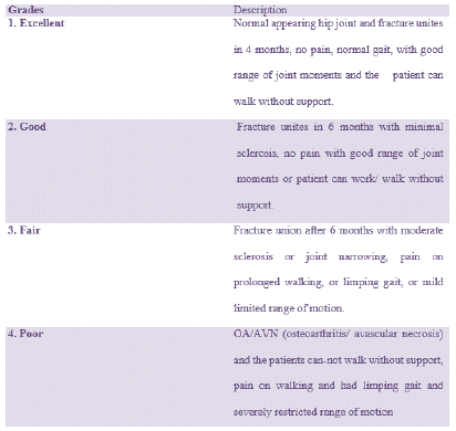
Patients were followed for a minimum of 6 months. Clinical grading was done according to D’aubigne and Postel modified by Matta [14]. Pain, gait and range of motion were assessed. Radiological grading was done on last visit according to Matta 14 criteria as: excellent (normal appearing hip joint), good (mild changes with minimal sclerosis and joint narrowing less than 1mm), fair (intermediate changes with moderate sclerosis and joint narrowing less than 50%), and poor (advanced changes). Both the clinical and radiological findings were calculated and the results were summed in Matta Grading [14] (Figure 1).

Figure 1: Overall survival, autologous stem cell transplant (ASCT) versus no ASCT (p=0.12).
Data was entered in specially designed proforma. Data was entered and analyzed by using SPSS version 22.0. Mean and standard deviation was calculated for quantitative variables like age and duration of injury. Frequency and percentage was calculated for categorical variables like gender and hip pain. Effect modifiers like age, gender and duration of injury was addressed through stratification of data. Post stratification chi square was applied. P value =0.05 was taken as statistical significant.
Results
There was total 21 patients having age range of 20-60 years with average age of 37.52 (Table 1). Male were 14 (66.7%) while female were 7 (33.3%), (Table 2). The cause of injury was road traffic accident in 13 (61.9%), Fall 6 (28.6%) and Physical Violence in 2(9.5%) patients (Table 3). Right side was involved in 13 (61.9%) while left was involved in 8 (38.1%) patients (Table 4). There was plate loosening in 2 (9.5%) cases (Table 5). Excellent results were obtained in 11 (52.4%), good in 7 (33.3%), fair in 2 (9.5%) and poor in 1 (4.8%) cases (Table 6).
    Age of the Patient


    N

    Valid

    21


    Missing

    0


    Mean

    37.52


    Std. Error of Mean

    2.3


    Median

    39


    Mode

    35


    Std. Deviation

    10.54


    Minimum

    20


    Maximum

    60
Table 1: Statistics.
    Frequency

    Percent

    Valid Percent

    Cumulative Percent


    Valid

    Female

    7

    33.3

    33.3

    33.3


    Male

    14

    66.7

    66.7

    100


    Total

    21

    100

    100
Table 2: Gender of patient.
    Frequency

    Percent

    Valid Percent

    Cumulative Percent


    Valid

    Fall

    6

    28.6

    28.6

    28.6


    Motor vehicle    accident

    13

    61.9

    61.9

    90.5


    Physical violence

    2

    9.5

    9.5

    100


    Total

    21

    100

    100
Table 3: Mechanism of trauma.
    Frequency

    Percent

    Valid Percent

    Cumulative Percent


    Valid

    Left

    8

    38.1

    38.1

    38.1


    Right

    13

    61.9

    61.9

    100


    Total

    21

    100

    100
Table 4: Side of involvement.
    Frequency

    Percent

    Valid Percent

    Cumulative Percent


    Valid

    No Problem

    19

    90.5

    90.5

    90.5


    Screw loosening

    2

    9.5

    9.5

    100


    Total

    21

    100

    100
Table 5: X-ray finding in first follow up.
    Frequency

    Percent

    Valid Percent

    Cumulative Percent


    Valid

    Excellent

    11

    52.4

    52.4

    52.4


    Fair

    2

    9.5

    9.5

    61.9


    Good

    7

    33.3

    33.3

    95.2


    Poor

    1

    4.8

    4.8

    100


    Total

    21

    100

    100
Table 6: Result in final follow up.
Discussion
Nowadays surgery is the corner stone of acetabular fracture management. Multiple approaches are used but the Kocher-Langenbeck and Ilioinguinal approaches remain the most common surgical approaches but the Anterior Intra-Pelvic approach has become relatively less common [10].
Open Rigid fixation of acetabulam fractures need expertise in the field of surgery and in old age patients it is a topic of debate because the results are different in may studies studies. Before going for surgery in old age patient the quality of bone and possibility of acceptable reduction should be kept in mind. A study done by Matta14 in which he reveiwed 255 acetabular fractures fixed by open reduction and internal fixation with the mean follow-up of 6 years [14]. He concluded that fracture reduction was the key for good clinical results and an increasing age of patient adversely affects the reduction as well as clinical results.
Tannast et al., has studied 816 patients that has been managed with fixation, showed that old age was a negative predictor of survivorship of hip joint [15]. He also proved that the there was poor outcome with anterior wall fracture of acetabulam.
There was 61% anatomical reduction in post-operative radiographs in Anglen et al., [16]. Study with ORIF of acetabular fractures in more than 60 years old patients. Some studies have reported poor outcome after acetabular fractures in elderly patients, reasonable outcome has been reported in other 7 patients after imperfect fracture reduction.
Miller et al., analysed post-operative fracture reduction after ORIF in 45 patients with mean age of 67 (range 59-82) years [17]. The authors achieved anatomical reduction in 26 patients only on post-operative radiographs, while none of the patients had anatomical reduction on CT scan. However, clinical results were not found to be correlated with radiological reduction at follow-up of average 72.4 months.
Similarly, Archdeacon et al., Concluded in their study that reasonable functional results can be achieved even without anatomical reduction of acetabular fractures in elderly patients [18]. The results of 18 patients have been analyzed by Helfet et al [19]. With an average age of 67 (range 60-81) years and concluded that open reduction and internal fixation of acetabular fracture in elderly patients can yield good outcomes.
Kelly J et al., Studied a total of 8389 acetabular fractures from 8372 patients [20]. The mean patient age was from 38.6 to 45.2. Road Traffic Accidents (RTA) accounting for 66.5% of cases and fractures caused by falls was 25.8%. A change in injury mechanisms is seen, with decrease in RTA, which was previously over 80%, and rise in the number of fall, which was previously 10.7%. He also notice a marked change in the pattern of type of fracture, with a significant rise in anterior column-based fractures (Anterior column and Anterior column posterior hemi-transverse), whilst all other fracture patterns have fallen over time. The most significant change in complications is a substantial drop in iatrogenic sciatic nerve injury. Post-traumatic osteoarthritis of the hip joint remains one of the most complication of acetabulam fracture, with 16.9% of cases developing Matta grade III/IV changes by 44 months in this review. Heterotopic ossification also remains a common problem [14].
Conclusion
Open reduction and internal fixation with 3.5 millimeter reconstruction plate and screws gives Excellent to Good results according to Matta Grading in all fracture of the acetabulam even in osteoporotic bone as well.
References
- Cimerman M, Kristan A, Jug M, Tomazevic M. Fractures of the acetabulum: from yesterday to tomorrow. Int Orthop. 2021; 45: 1057-1064.
- Pavelka T, Salasek M, Dzupa V. Priciny zmen spektra zlomenin acetabula v poslednich 20 letech [Causes of Changes in the Spectrum of Acetabular Fractures in the Last 20 Years]. Acta Chir Orthop Traumatol Cech. 2020; 87: 329-332.
- Mc Gee A, Obinwa C, White P, Cichos K, McGwin G, et al. Preoperative Blood Loss of Isolated Acetabular Fractures. J Orthop Trauma. 2023; 37: 116-121.
- Hartel MJ, Naji T, Fensky F, Henes FO, Thiesen DM, et al. The influence of bone quality on radiological outcome in 50 consecutive acetabular fractures treated with a pre-contoured anatomic suprapectineal plate. Arch Orthop Trauma Surg. 2022; 142: 1539-1546.
- Mansour A, Givens J, Whitaker JE, Carlson J, Hartley B. Immediate outcomes of early versus late definitive fixation of acetabular fractures: A narrative literature review. Injury. 2022; 53: 821-826.
- Johns BP, Balogh ZJ. The horizontal shear fracture of the pelvis. Eur J Trauma Emerg Surg. 2022; 48: 2265-2273.
- Chen G, Mo J, Zhang Y, Li R, Yang H, et al. Short-term effectiveness of reconstruction plate internal fixation via improved Stoppa approach combined with iliac fossa approach and Kocher-Langenbeck approach for complex acetabular fractures. Zhongguo Xiu Fu Chong Jian Wai Ke Za Zhi. 2022; 36: 1453-1458.
- Yao S, Chen K, Zhu F, Liu J, Wang Y, et al. Internal fixation of anterior acetabular fractures with a limited pararectus approach and the anatomical plates: preliminary results. BMC Musculoskeletal Disorder. 2021; 22: 203.
- Salameh M, Hammad M, Babikir E, Ahmed AF, George B, et al. The role of patient positioning on the outcome of acetabular fractures fixation through the Kocher-Langenbeck approach. Eur J Orthop Surg Traumatol. 2021; 31: 503-509.
- Shaath MK, Lim PK, Andrews R, Gausden EB, Mitchell PM, et al. Clinical Results of Acetabular Fracture Fixation Using a Focal Kocher-Langenbeck Approach Without a Specialty Traction Table. J Orthop Trauma. 2020; 34: 316-320.
- Patil A, Attarde DS, Haphiz A, Sancheti P, Shyam A. A Single Approach for Management of Fractures Involving Both Columns of the Acetabulum: A Case Series of 23 Patients. Strategies Trauma Limb Reconstr. 2021; 16: 152-160.
- Stavrakakis IM, Kritsotakis EI, Giannoudis PV, Kapsetakis P, Dimitriou R, et al. Sciatic nerve injury after acetabular fractures: a meta-analysis of incidence and outcomes. Eur J Trauma Emerg Surg. 2022; 48: 2639-2654.
- Liu X, Li M, Liu J, Liu Z, Zhang L, et al. Research progress of different surgical approaches in treatment of acetabular both-column fractures. Zhongguo Xiu Fu Chong Jian Wai Ke Za Zhi. 2021; 35: 661-666.
- Matta J.M. Fractures of the acetabulum: accuracy of reduction and clinical results in patients managed operatively within three weeks after the injury. J Bone Joint Surg Am. 1996; 78: 1632-1645.
- Tannast M, Najibi S, Matta J.M. Two to twenty-year survivorship of the hip in 810 patients with operatively treated acetabular fractures. J Bone Joint Surg Am. 2012; 94: 1559-1567.
- Anglen J.O., Burd T.A., Hendricks K.J., et al. The “Gull Sign”: a harbinger of failure for internal fixation of geriatric acetabular fractures. J Orthop Trauma. 2003; 17: 625-634.
- Miller A.N., Prasarn M.L., Lorich D.G., et al. The radiological evaluation of acetabular fractures in the elderly. J Bone Joint Surg Br. 2010; 92: 560-564.
- Archdeacon M.T., Kazemi N., Collinge C., et al. Treatment of protrusion fractures of the acetabulum in patients 70 years and older. J Orthop Trauma. 2013; 27: 256-261.
- Helfet DL, Borrelli J, Jr., Di Pasquale T, et al. Stabilization of acetabular fractures in elderly patients. J Bone Joint Surg Am. 1992; 74: 753-765.
- Kelly J, Ladurner A, Rickman M. Surgical management of acetabular fractures - A contemporary literature review. Injury. 2020; 51: 2267-2277.