Case Report
Austin J Clin Case Rep. 2023; 10(5): 1290.
Traumatic Abdominal Aortic Dissection, a Rare Near-Missed Diagnosis: A Case Report and Literature Review
Chadi Rahmani1,2*; Yassine Kherchttou1,2; Manal Rhezali1,2; Taoufik Abou Elhassan1,2; Hicham Nejmi1,2
1Anesthesiology, Intensive care and Emergency Department, University Hospital Mohammed VI of Marrakech, Morocco
2Faculty of Medicine and Pharmacy of Marrakech, Cadi Ayyad University, Morocco
*Corresponding author: Chadi Rahmani Anesthesiology, Intensive Care and Emergency department, University Hospital Mohammed VI of Marrakech, Morocco. Email: chadi.rahmani@gmail.com
Received: May 26, 2023 Accepted: June 21, 2023 Published: June 28, 2023
Abstract
Acute traumatic dissection of the abdominal aorta due to blunt trauma is a rare and life-threatening condition, often resulting in death. Immediate diagnosis is critical for successful treatment and improved prognosis. In this case presentation, a 60-year-old man was found unconscious after a road accident and was diagnosed with aortic dissection that went unnoticed initially due to associated severe injuries. CT-scan and ultrasound examination confirmed the diagnosis of abdominal aortic dissection. This case highlights the importance of a high index of suspicion when certain signs are present, such as seat-belt sign, bowel injury, or spinal injury. CT-scan is an accurate diagnostic tool, and close imaging follow-up is necessary as delayed complications can occur. Nonoperative and endovascular management is often chosen.
Keywords: Aortic injury; Abdominal aorta; Emergency; High-energy trauma; Computed tomography scan
Abbreviations: ICU: Intensive Care Unit; GCS: Glasgow Coma Scale; eFAST: Extended Focused Assessment with Sonography in Trauma; CT-Scan: Computed Tomography Scan; AAST: American Association for the Surgery of Trauma; TAAI: Traumatic Abdominal Aortic Injury; MRI: Magnetic Resonance Imaging
Introduction
Acute traumatic dissection of the abdominal aorta secondary to blunt trauma is a rare and life-threatening injury. These lesions may initially go unnoticed due to the simultaneous presence of other critical and more visible lesions. They are typically brought on by high-energy trauma that results in direct or indirect forces on the abdomen that are frequently associated with rapid deceleration [1]. Non-penetrating road crashes are responsible for about 90% of acute traumatic lesions. Following physical trauma, disruption of the intima, subintimal hemorrhage, and thrombosis are occasionally. Before medical assistance can be provided, the majority of acute traumatic injuries to the abdominal aorta are usually fatal. If the diagnosis can be made immediately, surgical treatment may be indicated.
We present a rare and life-threatening aortic dissection after blunt abdominal trauma, which was initially not detected due to technical insufficiency. This case highlights the importance of comprehending the nature of the trauma, its subsequent injuries, and the need for a body Computed Tomography Scan (CT-scan) with intravenous contrast enhancement in patients who have high-energy impacts.
Case Presentation
Patient Information
We present the case of a 60-year-old man, with no known (initially) identity or medical history, who was found unconscious in a likely traumatic context on the highway and transferred to the ICU by ambulance, the exact mechanism of the trauma was not known.
Clinical Findings
The primary assessment showed an unconscious patient with GCS of 3 with miosis progressed to anisocoria, parietal scalp hematoma and macroscopic hematuria, however the present patient was hemodynamically and respiratory stable, there was no auscultation abnormality. Abdominal clinical exam was normal, no hematoma was noted, and the examination of limbs was also normal with a normal distal pulse.
Diagnostic Assessment
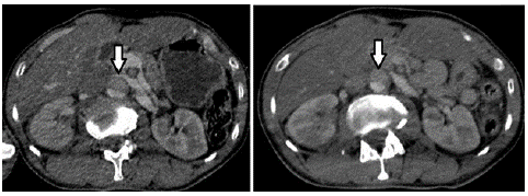
The eFast ultrasound performed showed no abnormalities. After initial conditioning, a body CT-scan was performed, revealing an acute subdural hematoma with a hypodense right frontal collection associated with subarachnoid and intraventricular hemorrhage (Figure 1). The abdominal CT-scan showed a perirenal hematoma associated with a 3cm lower polar kidney fracture, classified as AAST grade III, with an image of abdominal aortic thrombus (Figure 2).

Figure 1: Cerebral CT scan, revealing an acute subdural hematoma with a hypodense right frontal collection associated with subarachnoid and intraventricular hemorrhage.

Figure 2: Abdominal CT-scan showed a perirenal hematoma associated with a 3cm lower polar kidney fracture, classified as AAST grade III, with an image of abdominal aortic thrombus (Arrow).
Therapeutic Intervention
The patient was transported after discussion to the operating room for emergency acute subdural hematoma evacuation.
The surgery was performed without incident, the patient remained stable, pupils in miosis without signs of focusing.
Follow-up and Outcomes
In the postoperative period, the secondary study of the abdominal aortic thrombus picture prompted us to perform abdominal CT angiography. However, the images of the arterial phase were not optimal following a manual injection of the contrast product due to a technical error, a doubt was based on the presence of an intimal flap of the abdominal aorta (Figure 3).

Figure 3: Abdominal CT-scan showed the presence of an intimal flap of the abdominal aorta (Arrow).
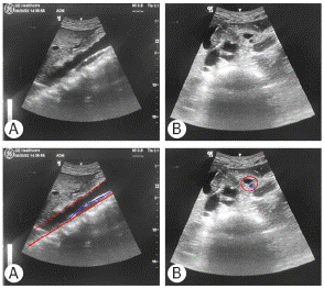
Moreover, an ultrasound examination at the patient's bedside revealed an intimal flap of the supra and sub renal abdominal aorta with partial thrombosis of the true channel (Figure 4), thus confirming the diagnosis of aortic dissection initially suspected on abdominal CT angiography. No intervention was performed and he was managed medically with a decision to perform, at day 3 if the situation remains stable, a control abdominal CT-scan to reevaluate the dissection and follow up on the aortic injury.

Figure 4: A) Ultrasound examination at the patient’s bedside B) longitudinal section C) Axial section D) revealed an intimal flap of the supra and sub renal abdominal aorta.
The evolution was fatal secondary to serious neurological lesions, marked initially by neurological worsening secondary to diffuse cerebral edema at the 4th postoperative hour, responsible for an areactive bilateral mydriasis resistant to neuroresuscitation and osmotherapy measures. Patient declared brain dead on the second postoperative day.
Discussion
Nonpenetrating traumatic injuries of the abdominal aorta remains rare, they are reported in less than 1% of blunt injuries. And only 25% of them result in aortic dissection. They are most commonly associated with high-speed vehicle collisions or other circumstances such as being run over or falling from a height building [1].
The mechanism of injury can result from direct or indirect impact damage. Direct impact damage always described in the context of seat belt syndrome, where the impact is believed to produce compression of the aorta against the vertebrae [2]. Indirect impact damage is caused by Shear forces which are an important factor in thoracic aortic injuries with a less important role in abdominal aortic injuries, where sudden acceleration can cause lesions mainly in the intima, or the increase in intraluminal pressure which must reach very high levels (more than 1000 mmHg) to cause injury, so this mechanism is observed less frequently [3]. In our case, the exact mechanism was unknown, which shows the interest of keeping in mind the possibility of aortic injury in any abdominal trauma.
Due to the nature of the mechanism and the strong forces required to injure the aorta, there are often accompanying injuries [4]. In our case, it was a lower polar kidney fracture with perirenal hematoma. Different injury patterns have been described, and the most common injuries associated with seat-belt injuries to the aorta are small-bowel injuries and flexion-distraction injuries of the lumbar spine. Others traumatic injuries like hemoperitoneum, solid organ injury, mesenteric hematoma, and colon injury are often observed [1,4].
A high degree of suspicion for aortic injury must be maintained in patients presenting with blunt abdominal trauma if they have associated injuries including a Chance vertebral fracture, one of the most common accompanying injuries in this context. Bowel injury, a “seat-belt sign,” or neurovascular deficits [2].
The clinical presentation of Traumatic Abdominal Aortic Injury (TAAI) depends on the type and severity of injury. In severe cases, patients may present with a hypovolemic shock (due to acute bleeding and retroperitoneal hematoma), acute abdomen and hemodynamic abnormalities. In these cases, physical examination may show signs of peritonitis and a weak, delayed, or absent pulse in the lower extremities. In less severe cases, like this one, there may be no abnormality in the clinical presentation and TAAI may be detected incidentally on imaging [5].
Historically, arteriography was considered the gold standard for diagnosis of aortic injuries. The dissection is mostly localized in the infrarenal aorta [1]. Emergency physicians routinely use bedside ultrasound to evaluate the abdominal aorta for aneurysm. Visualization of an intimal flap by ultrasound may carry a sensitivity of 67–80% and specificity of 99–100% for dissection [6]. This rapid, non-invasive method of diagnosis may aid in the early detection and treatment of this deadly diagnosis.
Modern CT has become the imaging method of choice in assessing the trauma patient and is now an integral part of injury assessment. Sagittal and coronal reformatted images can be used to detect subtle lesions such as intimal defects. Dual CT also promises to be useful as an advanced technique for early detection of vascular lesions [7]. However, some works relate that the diagnosis was made during surgery in 10% of cases [8], and only indirect signs are visible in the CT scan such as the presence of a retroperitoneal hematoma or strands of fat around the aorta.
The classification and treatment of aortic trauma is usually based on the traumatic experience of the thoracic aorta, but the same classification has been used in the literature for the abdominal aorta [9]. There have been various classification proposals, the most common being that of Azzizadeh et al., which divides the pathology into four categories according to imaging techniques: intimal tear (grade I), intramural hematoma (grade II), aortic pseudoaneurysm (grade III), and free rupture (grade IV) [10]. The goal of classifying lesions by imaging is to determine their severity and prognosis in order to decide which course of action is most appropriate: immediate treatment, delayed treatment, or follow-up. We believe that rapid and accurate description of imaging agents is essential in aortic lesions, since grade IV aortic lesions and most pseudoaneurysms require immediate treatment, and the rest of the lesions are likely to require reassessment or treatment in the future.
Many options are available for the treatment of the described injury, but the current data of the literature suggest treatment by stenting if feasible or by surgical replacement in selected cases. In the absence of ischemic paraplegia or other injuries that require emergency surgery, endovascular treatment is a safe and efficient method for treating traumatic infrarenal aortic dissection. Endovascular therapy appears to be associated with a relatively low risk of mortality or major complications compared to open repair and conservative treatment [8].
Conclusions
In summary, abdominal aortic dissection is a rare but potentially lethal complication of abdominal trauma and can occur at essentially any age. A high index of suspicion is warranted when a seat-belt sign, bowel injury, or flexion distraction spinal injury is present. CT-scan is accurate in its diagnosis, but this injury can be seen on MRI, ultrasound, and angiography. Management decisions are based on the individual patient, but nonoperative management is often chosen for the hemodynamically stable patients. Close imaging follow-up is mandatory because delayed complications can occur.
Author Statements
Declaration of Conflicting Interests
The authors declared no potential conflicts of interest with respect to the research, authorship, and/or publication of this article.
Consent for Publication
Written informed consent was obtained from the family patient for publication of this case report and any accompanying images. A copy of the written consent is available for review by the Editor-in-Chief of this journal.
Availability of Supporting Data
All clinical finding and radiological results included in this case report can be found in the archived medical file of the patient.
Acknowledgments
The authors would like to thank the team of the department of radiology, University Hospital Mohammed VI of Marrakech for the great work they do and for their essential role in this case report.
Funding
The authors received no financial support for the research, authorship, and/or publication of this article.
Authors' Contributions
All authors have contributed to this work since conception, reading and endorsing the final version of the manuscript.
References
- Prado E, Chamorro EM, Marín A, Fuentes CG, Zhou ZC. CT features of blunt abdominal aortic injury: an infrequent but life-threatening event. Emerg Radiol. 2022; 29: 187–195.
- Lalancette M, Scalabrini B, Martinet O. Seat-belt aorta: a rare injury associated with blunt abdominal trauma. Ann Vasc Surg. 2006; 20: 681–683.
- Baghdanian AH, Armetta AS, Baghdanian AA, LeBedis CA, Anderson SW, et al. CT of Major Vascular Injury in Blunt Abdominopelvic Trauma. Radiogr Rev Publ Radiol Soc N Am Inc. 2016; 36: 872–890.
- Macura KJ, Corl FM, Fishman EK, Bluemke DA. Pathogenesis in acute aortic syndromes: aortic dissection, intramural hematoma, and penetrating atherosclerotic aortic ulcer. AJR Am J Roentgenol. 2003; 181: 309–316.
- Gunn M, Campbell M, Hoffer EK. Traumatic abdominal aortic injury treated by endovascular stent placement. Emerg Radiol. 2007; 13: 329–331.
- Fojtik JP, Costantino TG, Dean AJ. The diagnosis of aortic dissection by emergency medicine ultrasound. J Emerg Med. 2007; 32: 191–196.
- Wortman JR, Uyeda JW, Fulwadhva UP, Sodickson AD. Dual-Energy CT for Abdominal and Pelvic Trauma. Radiogr Rev Publ Radiol Soc N Am Inc. 2018; 38: 586–602.
- Jonker FHW, Schlösser FJV, Moll FL, Muhs BE. Dissection of the abdominal aorta. Current evidence and implications for treatment strategies: a review and meta-analysis of 92 patients. J Endovasc Ther Off J Int Soc Endovasc Spec. 2009; 16: 71–80.
- Heneghan RE, Aarabi S, Quiroga E, Gunn ML, Singh N, et al. Call for a new classification system and treatment strategy in blunt aortic injury. J Vasc Surg. 2016; 64: 171–176.
- Azizzadeh A, Keyhani K, Miller CC, Coogan SM, Safi HJ, et al. Blunt traumatic aortic injury: initial experience with endovascular repair. J Vasc Surg. 2009; 49: 1403–1408.