Special Article: Radiology Case Reports
Austin J Clin Case Rep. 2023; 10(6): 1296.
Responsibility Comes with the Dominance: A Rare Case Report of Absent Right Coronary Artery with Super Dominant Left Circumflex Artery
Kritisha Rajlawot¹*; Nirmal Prasad Neupane¹; Manisha Aryal¹; Dharmanath Yadav²
¹Radiologist, Department of Radiodiagnosis and Imaging, Shahid Gangalal National Heart Center, Bansbari, Kathmandu, Nepal
²Cardiologist, Department of Cardiology, Shahid Gangalal National Heart Center, Bansbari, Kathmandu, Nepal
*Corresponding author: Kritisha Rajlawot Radiologist, Department of Radiodiagnosis and Imaging, Shahid Gangalal National Heart Center, Bansbari, Kathmandu, Nepal. Tel: +9779849928166 Email: nirmal.neup@gmail.com
Received: June 21, 2023 Accepted: August 01, 2023 Published: August 08, 2023
Abstract
Coronary artery dominance describes the source from which the Posterior Descending Artery (PDA) is arising that supplies the inferior wall. 80-85% of the times it is Right Coronary Artery (RCA) referring to right dominance, 7-13% of the times it is Left Circumflex artery (LCx) referring to left dominance and about 2-5% of the times it is originating from both the RCA and LCx making a codominant supply. A super dominant vessel is when a vessel is exceptionally huge supplying the area that the other vessel usually covers which makes cardiac circulation rely on one vessel. Here we present a rare case of absent RCA with super dominant LCx supplying the RCA territory diagnosed through Coronary Computed Tomography Angiography (CCTA). As invasive coronary angiography may not always provide sufficient information in such cases, CCTA is considered as a robust, reliable and non-invasive modality of imaging to investigate so as to avoid an ischemic insult.
Keywords: Coronary computed tomography angiography (CCTA); Absent right coronary artery; Super dominant left circumflex artery; Invasive coronary angiography
Introduction
Coronary artery dominance is simply described by the source from which the Posterior Descending Artery (PDA) is arising and supplying the inferior wall or the posterior interventricular septum. 80-85% of the times it is Right Coronary Artery (RCA) referring to right dominance, 7-13% of the times it is Left Circumflex artery (LCx) referring to left dominance and about 2-5% of the times it is originating from both the RCA and LCx making a codominant supply [1]. Amongst various presentation of coronary artery dominance, a super dominant vessel is when a vessel is exceptionally huge supplying the area that the other vessel usually covers. Different variations of the super dominant coronaries have been reported in the literature previously such as the territory of the RCA as well as the Left Anterior Descending (LAD) artery being supplied by a super-dominant LCx artery [2], super dominant RCA extending along the base of the heart to left atrioventricular groove where it is supplying the lateral wall of the left ventricle and LCx being completely absent, with no visible vessel in the upper portion of the left atrioventricular groove [3], super dominant RCA with double Posterior Descending Artery (PDA) [4], super dominant LAD supplying the apex and giving rise to the posterior interventricular artery, that is ascending along the groove until the crux cordis with continuation as a posterior lateral branch [5]. As being mentioned the super dominant vessel may present in several forms which is yet an uncommon form of anomalous coronary with an incidence of less than 0.066% [6]. As it may not have a typical clinical presentation, super dominant vessel or congenital absence of one of the coronaries or two is not a first diagnosis that comes to anyone’s mind with a symptom of acute chest pain. Therefore, here we attempt to present a rare case of absent RCA with super dominant LCx supplying the RCA territory.
Clinical History
A female patient aged 70 presented to our out-patient department with complaints of chest pain, associated with shortness of breath that relieves on rest and associated palpitation. She had a history of diabetes, hypertension and hyperlipidemia under medications. Her electrocardiography showed ST segment changes, while her cardiac enzymes were negative. Her treadmill test was however positive. Therefore, with a provisional diagnosis of stable angina at moderate efforts, she was planned for an elective invasive coronary angiography considering her age and comorbidities. During the procedure, RCA was not well visualized and hence the patient was referred to undergo Coronary Computed Tomography Angiography (CCTA) for further evaluation so as to rule out an obstructed proximal RCA or an anomaly or a dominant LCx.
Imaging Features
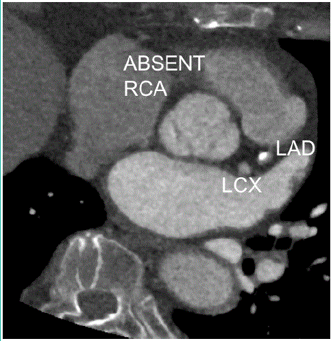
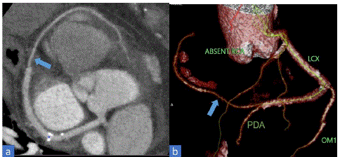
The patient underwent a CCTA for the evaluation of the RCA and also to rule out significant stenoses of the coronaries. There was normal origin of the left main artery from the left sinus of valsalva that was further branching into LAD and LCx arteries. The calcium scoring was significantly higher corresponding to above 800 agatston units where LAD was diffusely diseased showing about 70-80% luminal stenosis. LCx also had eccentric calcified plaques with about 20-30% luminal stenosis. However, the origin of RCA from the right sinus of Valsalva was not appreciated with an absent ostium and proximal segment (Figure 1). On the other hand, a large LCx was noted extending into the right atrioventricular groove and terminating near the right sinus of Valsalva perfusing the RCA territory (Figure 2). Clearly LCx was a dominant vessel as it was giving off the Posterior Descending Artery (PDA) (Figure 2b). Hence, a diagnosis of absent RCA with super dominant LCx supplying the RCA territory was given.

Figure 1: Coronary computed tomography angiography axial view showing normal branching of left main artery into Left Anterior Descending (LAD) and Left Circumflex (LCx) artery. Absent Right Coronary Artery (RCA) from the right sinus of Valsalva.

Figure 2: (a) Coronary computed tomography angiography axial view and (b) 3D volume-rendered coronary image showing Left Circumflex artery (LCx) extending to the right atrioventricular groove (arrow) supplying the Right Coronary Artery (RCA) territory.
Discussion
The occurrence of super dominant vessel as in our case has been reported as anomalous RCA coming from the LCx which corresponds to left type I (L-I type) variant of single coronary artery. According to Lipton-Yamanaka classification of single coronary artery, variations of single coronary artery was introduced based on the origin and course in relation to the ascending aorta and pulmonary trunk. Nevertheless, the incidence of the L-I subtype was only 0.016% [7-10]. Although the L-I subtype is generally regarded as benign and typically treated medically, the non-visualization of the RCA from the right coronary sinus in the setting of acute coronary syndromes might be mistaken for an ostial RCA occlusion. Any attempt at revascularization may cause unintentional harm, for instance perforation of the coronary sinus due to aggressive manipulation of the guidewire [11]. Hence, upon non-visualization of any coronary artery, coronary anomalies in the form of absent coronary artery with super dominant vessel or single coronary artery needs to be considered. The coronary anomalies are rare finding discovered incidentally while undergoing coronary angiogram. Any type of coronary anomaly is important to be timely diagnosed and managed if diseased, as stenosis of the dominant vessel can risk a large portion of the myocardium, which if left untreated can lead to fatal outcomes [10]. CCTA with steadily improved high spatial and temporal resolution essential for imaging the coronaries, has gradually been established as an accurate and reliable mode of imaging coronary arteries. Although invasive coronary angiogram is considered the clinical gold standard for evaluating the coronary arteries, it has numerous drawbacks, first being an invasive procedure it carries a small risk of morbidity and mortality. Second, its projectional limits may cause difficulty to precisely characterize the anatomical variants, especially in the setting of coronary abnormalities. Therefore, there are list of conditions where non- invasive CCTA is preferred over invasive coronary angiography, for example: in ruling out significant luminal stenosis in stable but suspicious cases of coronary stenoses, to rule out coronary artery disease in patients presenting with acute chest pain, a pre-operative investigation of choice to rule out coronary stenoses, to evaluate patency of bypass grafts and stents, and undoubtedly to assess the variabilities of coronary anomalies [12-15]. Hence, CCTA has been embraced as a promising imaging option for additional study of the intricate and challenging anatomical differences. Patients with such coronary anomalies are treated based on the clinical symptoms, where each variety is managed using a different approach including conservative treatment, or stent placement, or even surgical correction if required. In our case, after being given an impression of absent RCA with super dominant LCx, the patient underwent stenting for diffusely diseased LAD with additional medical management for non-critical plaques.
Conclusion
The presence of super dominant vessel in an individual makes cardiac circulation rely on one vessel, which might cause the enormous repercussions in cases of significant occlusion. Therefore, it is crucial to recognize the clinical significance of such rare entity of having super-dominant vessel associated with absence of another major vessel. At the same time, it is important to choose the appropriate choice of imaging to evaluate the super dominant vessel that may exist in different forms of single coronary artery. Certainly, invasive coronary angiography may not always provide sufficient information in such cases, hence making CCTA a robust, reliable and non-invasive modality of imaging to investigate so as to avoid an ischemic insult.
Author Statements
Competing Interests
All of the authors declare that they have no competing interests.
Ethics Approval and Consent to Participate
This case report did not require review by the Ethical committee for publication.
Funding
No funding was obtained for this study.
References
- Aricatt DP, Prabhu A, Avadhani R, Subramanyam K, Manzil AS, Ezhilan J, et al. A study of coronary dominance and its clinical significance. Folia Morphol (Warsz). 2023; 82: 102-7.
- Agrawal N. Superdominant left-circumflex artery supplying significant proportion of RCA and LAD territory. BMJ Case Rep. 2015; 2015: bcr2015210365.
- Quijada-Fumero A, Pimienta-González R, Rodriguez-Esteban M. Absence of left circumflex with superdominant right coronary artery. BMJ Case Rep. 2014; 2014: bcr2014206782.
- Maheshwari M, Mittal SR. Superdominant right coronary artery with double posterior descending artery. Heart Views. 2015; 16: 19-20.
- Rodríguez-Blanco S, Valdés-Recarey M, Leyva Quert AY. Super dominant left anterior descending coronary artery. Rev Esp Cardiol (Engl Ed). 2017; 70: 1006.
- Canan A, Batra K. Superdominant left circumflex artery with absent right coronary artery. Radiology. 2022; 304: 294.
- Lipton MJ, Barry WH, Obrez I, Silverman JF, Wexler L. Isolated single coronary artery: diagnosis, angiographic classification, and clinical significance. Radiology. 1979; 130: 39-47.
- Yamanaka O, Hobbs RE. Coronary artery anomalies in 126,595 patients undergoing coronary arteriography. Cathet Cardiovasc Diagn. 1990; 21: 28-40.
- Chou LP, Kao C, Lee MC, Lin SL. Right coronary artery originating from distal left circumflex artery in a patient with an unusual type of isolated single coronary artery. Jpn Heart J. 2004; 45: 337-42.
- Iftikhar I, Rehman AU, Khan HS. A single coronary artery with an absent right coronary and a superdominant left circumflex giving off a branch supplying the right coronary artery territory. J Coll Physicians Surg Pak. 2019; 29: S80-2.
- Oliveira MDP, Navarro EC, Ferraz TX, Silveira FS, de Sá GA, Castello Júnior HJ et al. Superdominant left circumflex with absence of the right coronary artery: an interesting and very rare coronary anomaly. J Xiangya Med. Dec 2018; 3: 42.
- Vanhoenacker PK, Heijenbrok-Kal MH, Van Heste R, Decramer I, Van Hoe LR, Wijns W et al. Diagnostic performance of multidetector CT angiography for assessment of coronary artery disease: meta-analysis. Radiology. 2007; 244: 419-28.
- Hoffmann U, Nagurney JT, Moselewski F, Pena A, Ferencik M, Chae CU et al. Coronary multidetector computed tomography in the assessment of patients with acute chest pain. Circulation. 2006; 114: 2251-60.
- Nieman K, Pattynama PM, Rensing BJ, Van Geuns RJ, De Feyter PJ. Evaluation of patients after coronary artery bypass surgery: CT angiographic assessment of grafts and coronary arteries. Radiology. 2003; 229: 749-56.
- Dodd JD, Ferencik M, Liberthson RR, Cury RC, Hoffmann U, Brady TJ et al. Congenital anomalies of coronary artery origin in adults: 64-MDCT appearance. AJR Am J Roentgenol. 2007; 188: W138-46.