Special Issue: Veterinary Case Reports
Austin J Clin Case Rep. 2023; 10(6): 1299.
Assessment and Non-Bandaging Treatment of Third-degree Thermal Injury in the Dog: A Case Report
Kuclar Muftic S; Lutvikadic I*; Maksimovic A
¹Clinical Department, University of Sarajevo – Veterinary Faculty, 71000 Sarajevo, Bosnia and Herzegovina
*Corresponding author: Lutvikadic I Clinical Department, University of Sarajevo – Veterinary Faculty, 71000 Sarajevo, Bosnia and Herzegovina Tel: +387 60 342 0137 Email: ismar.lutvikadic@vfs.unsa.ba
Received: July 14, 2023 Accepted: August 31, 2023 Published: Septbember 07, 2023
Abstract
Dogs– scalds are a significant concern in veterinary practice. The causes can vary with common sources including hot water, boiling liquids, heated surfaces, and household accidents. Factors such as age, breed, and environment may influence the likelihood of scald injuries in dogs. Human negligence can contribute to the occurrence of scald injuries therefore pet owners should be educated about potential hazards in the household to ensure a safe environment and provide proper supervision and significantly reduce the risk of scald injuries. Thermal burns cause a spectrum of injuries, ranging from superficial to deep, leading to local damage and potential systemic effects based on the severity. It is imperative that owners understand importance of the early pet evaluation by professionals, or to provide first aid. Accurate clinical estimation is crucial for effective treatment and prognosis. Throughout history, the calculation of the affected body surface area of the animal was based on different methods in human medicine. Recent studies showed this method as inaccurate and new calculations were proposed. Because of the wide diversity, a completely accurate method is not established in veterinary medicine yet. Although standard treatment of burns is not established in veterinary medicine, the administration of analgesics, antibiotics, and fluid therapy is frequently recommended as essential. To stimulate wound healing and prevent excessive pain, wound moisturize is recommended as a part of treatment. We report the assessment and treatment of four days old third-degree thermal burn injury in an adult Pekingese dog.
Keywords: Burn; Dog; Emergency; Thermal injury; Scalds
Introduction
Burns are injuries with extensive lesions of the skin mostly caused by high temperatures. Depending on the severity, subcutaneous and muscle tissue can be affected which can result in many metabolic complications [1,2]. The general condition of patients presented with burns depends on the temperature applied to the tissue, the duration of exposure, the type of heat transfer, and other factors [3]. Two basic parameters of burns are the degree of injury and the percentage of the affected area [1,4]. Clinically, in order to provide the best possible therapeutic plan and adequate prognostic assessment, it is very important to assess the Burn Surface Area (BSA). According to the CT results from the study by Henriksson et al. [5], it is estimated that the head and abdomen cover 14%, the neck and individual thoracic limbs 9%, the thorax 18%, the pelvic limbs individually 11%, and the pelvis and tail covers 5% of the total body surface in mesocephalic dogs. Due to wide diversity, there is no accurate method for BSA estimation in animals [6]. Furthermore, classifying burns is a crucial aspect of patient evaluation. Depending on the degree of injury it can be classified as first-, second-, third-, and fourth-degree burns [2,7]. In many cases, thermal injuries are treated as traumatic wounds [1]. After complete triage of the patient and clinical examination, it is necessary to start with immediate treatment which depends on the injury type [6]. It is very important to keep the affected area moist in order to prevent additional irritation of the nerve endings which will reduce the pain [8]. For this purpose, it is preferable to bandage the burn with non-adherent bandages by applying different topical mixtures [2,6]. Only in cases of difficult bandaging, the burn wound can remain exposed to atmospheric air with maximum care and prevention of secondary infections [1]. We described the management of a third-degree, wide-body surface thermal burn in an adult Pekingese dog.
Case Description
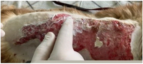
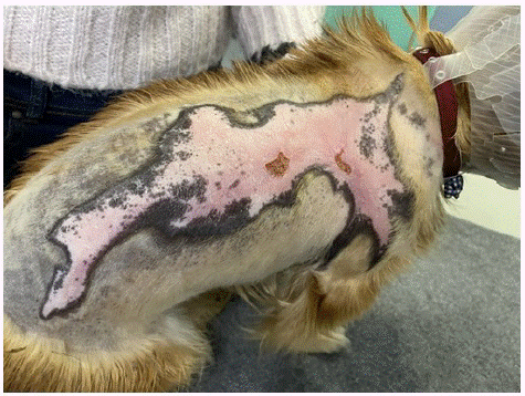
A four years old male Pekingese dog was presented to the Clinic for Surgery, Anesthesia and Resuscitation of the University of Sarajevo - Veterinary Faculty. The dog's injury was caused by boiling water, four days prior to admission. During this period, first aid or emergency treatment was not initiated. The owners noticed increased aggression and changes in the appearance of hair on the right side of the thorax. No changes in mental state were observed during the clinical examination, except for aggression regardless of extremely careful physical examination. The area of stuck and burnt hair with underneath skin damage was visible in the region of the right thorax (Figure 1). The dog was sedated by intramuscular administration of a combination of medetomidine hydrochloride (0.01 mg·kg–1) (Sedastart 1mg/ml, Dechra, UK) and methadone (0.3 mg·kg-1) (Comfortan 10 mg·mL-1, Dechra, UK). After achieving an adequate level of sedation, intravenous cannulation was performed with a 24G catheter (Mediplus Limited, India) on v. cephalica and the racemic mixture of propofol (2 mg·kg-1) (Propofol Claris 10mg/ml, SanMed GmbH, Austria) and ketamine hydrochloride (2 mg·kg-1) (Ketaminol10 100mg/ml, MSD, Netherland) was administered intravenously to induce anesthesia. In our case, we opted for general anesthesia with ketamine in addition to achieving better analgesic effects in combination with opioids. The estimated dehydration of the dog was 6%, subjectively based on physical presentation. No signs of malnutrition were reported and supportive feeding was not necessary. The Hartmann's solution in a dose obtained from the formula of 2 mg·kg-1 x %BSA during the first 24h and CRI with fentanyl (Fentanyl 0.05 mg·mL-1, Panpharma S.A., France) at a dose of 3 mcg·kg-1·h-1 was initiated. Immediately after establishing complete immobilization of the patient, endotracheal intubation was performed for the patient's oxygenation. Basic anesthesia monitoring was performed recording the results of ECG, respiration, and SpO2 by multiparameter anesthesia device (Mindray iMEC8Vet, China). After assessing the anesthesia depth, the removal of the hair and a more detailed examination of the body surface and wound were performed. Due to affected epidermis layers and tissue necrosis, the injury was classified as third-degree burn. The injury was observed on the right side of the thorax, right front limb, and abdomen, extending from the right shoulder joint to the right inguinal region. After mechanically removing the necrotic surface layer, the remaining tissue structures showed satisfactory tissue quality with a BSA estimate of more than 20%. Treatment with silver-sulfadiazine cream (Dermazin 1%, Sandoz, Croatia) was started after wound debridement (Figure 2), and the patient was hospitalized next 24 hours. The recommended treatment after discharge was based on the oral administration of carprofen (3 mg·kg-1) (Rycarfa 20mg, Krka, Croatia) and tramadol (2 mg·kg-1) (Boldol 50mg, Bosnia and Herzegovina) for the next 10 days, keeping the wound surface moist using a thin layer of silver-sulfadiazine cream every 8 hours. The use of the Elizabethan collar was also a mandatory instruction for the patient's owners to prevent further tissue damage and irritation. Analgesic management showed a satisfactory effect, which resulted in a positive change in the dog's behavior toward the owners and aggressive behavior disappeared. Mechanical debridement was performed only once on admission and such treatment was no need to repeat. Three weeks after the initial treatment and regular visitations, the epithelization of the wound was completed (Figure 3). None of systemic disorders were detected by laboratory and clinical results during the regular visitations.

Figure 1: Overall survival, autologous stem cell transplant (ASCT) versus no ASCT (p=0.12).

Figure 2: Application of silver-sulfadiazine cream after the wound cleaning in general anesthesia.

Figure 3: Complete wound epithelization 22 days after initial treatment.
Discussion
In human and veterinary medicine, burns represent a significant issue ranking third among all injuries [9]. There are several different types of burns, but thermal injuries are the most common with an incidence of 86% [4,10]. It is very important to do a proper estimation of the burn in order to provide the best clinical management. To achieve a correct estimation of the injury, the depth of the burn and the percentage of the affected area should be calculated [1,4]. In human medicine, several methods are available to calculate affected BSA, whereas the “Lund-Browder Chart” is considered the most precise method [4]. Historically, many of these methods were used in veterinary medicine but they are inaccurate [11,12]. Using the CT, Henriksson et al. [5] suggested body mapping in mesocephalic dogs. That is, they gave the percentage representation of the body surface through specific individual parts. Results showed that the head and abdomen cover 14%, the neck and individual thoracic limbs 19%, the thorax 18%, the pelvic limbs individually 11%, and the pelvis and tail cover 5% of the body surface [5]. Comparing these suggestions with methods in human medicine, inaccuracy is evident in earlier methods. BSA mapping is crucial considering that burns covering more than 10% of the body can be life-threatening causing severe changes in the animal's body, whereas burns covering more than 20% BSA in human medicine are classified as severe [2,4,7]. However, all burn injuries which do not extend 40% BSA may have a favorable treatment prognosis [6].
For successful treatment, it is imperative to start management as soon as possible after the injury. Burns treatment mainly depends on the depth of the injury but in veterinary medicine unique, standardized treatment is not established [2,6]. Generally, treatment should start with analgesics, antimicrobials to prevent infection, fluid therapy for patients with more than 20% BSA affected, and local treatment with silver-sulfadiazine cream as a first choice [13]. Nowadays, it is well known that systemic antimicrobials are not effective in infection prevention of susceptible tissue. Even though the necrotic tissue in third-degree burns is a good breeding ground for microorganisms [2], antimicrobial treatment of our patient was based only on local therapy. Furthermore, it is very important to prevent nerve receptors dehydration which can lead to additional pain [14]. This can be achieved by wet bandages with different drugs or drug combinations. Bandaging treatment in burned animals is often recommended and it showed a faster healing according to recent studies [2]. As the moist enhance wound healing and analgesic effect [8,15], our treatment goal was primarily based to accomplish this requirement. To achieve frequent wound care with less nerve-ending irritation, we decided to keep the affected area uncovered. Subjectively, such a decision showed a faster healing process with a better outcome for the animal due to better cooperativeness and diminishes aggression previously present because of pain. A combination of the drugs with analgesic properties, regular local moisturizing, and general care resulted in progressive favorable behavior change. In our case, non-bandaging method without systemic use of antimicrobials was sufficient.
Conclusion
In conclusion, achieving the best outcome in treating thermal injuries in veterinary patients requires an accurate calculation of the BSA using the most precise veterinary method available, correct assessment of the tissue injury extent and classification of the burn degree, and to identified and prevent any potential local and/or systemic complications. Only with a comprehensive clinical approach to burn injuries appropriate management can be ensured, leading to a favorable prognosis.
References
- Garzotto CK. Thermal burn injury. In: Silverstein DC, Hopper K, editors. Small animal critical care medicine. 2009; 2009: 683-6.
- Shnyakina TN, Shcherbakov NP, Bryukhanchikova NM, Medvedeva LV, Bezin AN. Experience in treatment of thermal burns in dogs. E3S Web Conf. 2021; 282: 03021.
- Bohnert M. Injury: burns, scalds, and chemical. In: Jamieson A, Moenssens AA, editors. Wiley encyclopedia of forensic science; 2009.
- Schaefer TJ, Szymanski KD. Burn evaluation and management. Available from: https://www.ncbi.nlm.nih.gov/books/NBK430741/. In: Stat pearls, Treasure Island (FL); 2022.
- Henriksson A, Kuo K, Gerken K, Cline K, Hespel AM, Cole R, et al. Body mapping chart for estimation of percentage of body surface area in mesocephalic dogs. J Vet Emerg Crit Care (San Antonio). 2022; 32: 350-5.
- Bolcato M, Roccaro M, Gentile A, Peli A. First report on medical treatment and outcome of burnt cattle. Vet Sci. 2023; 10: 187.
- Efimova OI, Dimitrieva AI, Nesterova OP, Aldyakov AV, Obukhova AV, Ivanova TN. Methods for the effective treatment of animal burns. IOP Conf Ser Earth Environ Sci. 2019; 346: 012057.
- Fowler A. Recognition of pain in wildlife. Wildcare Australia. 2007; 44: 8-11.
- Bezina NM. Clinical and hematological status of dogs in the treatment of burn injury. Materials of the International Scientific and Practical Conference: Veterinary medicine – to the agro-industrial complex. Toritsk. 2017; 35-9.
- Domaszewska-Szostek AP, Krzyzanowska MO, Czarnecka AM, Siemionow M. Local treatment of burns with cell-based therapies tested in clinical studies. J Clin Med. 2021; 10: 396.
- Vaughn L, Beckel N, Walters P. Severe burn injury, burn shock, and smoke inhalation injury in small animals. Part 2: Diagnosis, therapy, complications, and prognosis. J Vet Emerg Crit Care (San Antonio). 2012; 22: 187-200.
- Vigani A, Culler CA. Systemic and local management of burn wounds. Vet Clin North Am Small Anim Pract. 2017; 47: 1149-63.
- Dahlqvist E, Lindford A, Koljonen V. Conservative treatment of a scald in a puppy. EPlasty. 2009; 9: e46.
- Fleck CA. Wound pain: assessment and Management. Clin Pract. 2007; 5: 10-6.
- Alshehabat M, Hananeh W, Ismail ZB, Rmilah SA, Abeeleh MA. Wound healing in immunocompromised dogs: A comparison between the healing effects of moist exposed burn ointment and honey. Vet World. 2020; 13: 2793-7.