Case Report
Austin J Clin Case Rep. 2023; 10(8): 1309.
ST-Elevation Myocardial Infarction in Patient with Subarachnoid Hemorrhage
Grabczewska Z¹; Serafin Z²; Sciesinski J²
¹Department of Cardiology, Collegium Medicum in Bydgoszcz, Nicolaus Copernicus University in Torun, Poland
²Department of Radiology and Diagnostic Imaging, Collegium Medicum in Bydgoszcz, Nicolaus Copernicus University in Torun, Poland
*Corresponding author: Zofia Grabczewska Department of Cardiology, Antoni Jurasz University Hospital No. 1 ul. Marii Sklodowskiej-Curie 9, 85-094 Bydgoszcz Email: z.grabczewska@cm.umk.pl
Received: October 20, 2023 Accepted: November 13, 2023 Published: November 20, 2023
Abstract
We present the case of a 35-year-old man with ST-elevation lateral wall myocardial infarction and Subarachnoid Hemorrhage (SAH) from a ruptured aneurysm of the Anterior Cerebral Artery (ACA). Before the stroke was diagnosed, Electrocardiography (ECG) was performed, which showed changes characteristic of lateral wall ST-elevation myocardial infarction. Therefore, coronary angiography was carried out, revealing no stenotic lesions in the coronary arteries. Myocardial necrosis was confirmed by high troponin I levels and akinesia of lateral wall segments found in echocardiography examination. Once SAH was diagnosed, percutaneous embolization of the aneurysm was performed. Despite all the medical interventions undertaken, the patient died.
Key words: subarachnoid hemorrhage, myocardial infarction
Main Text
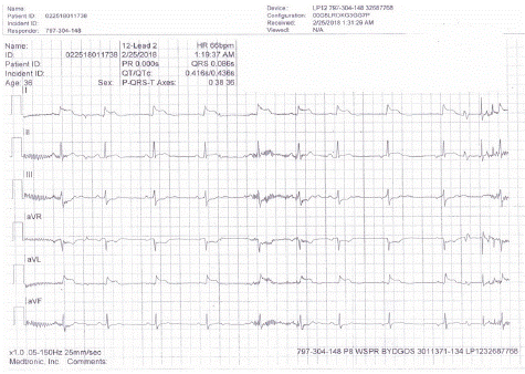
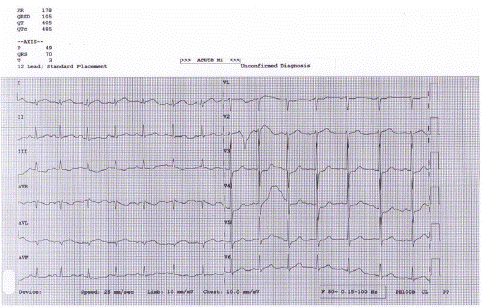
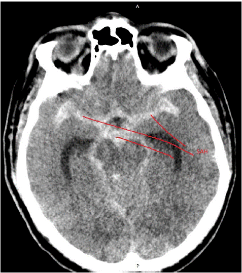
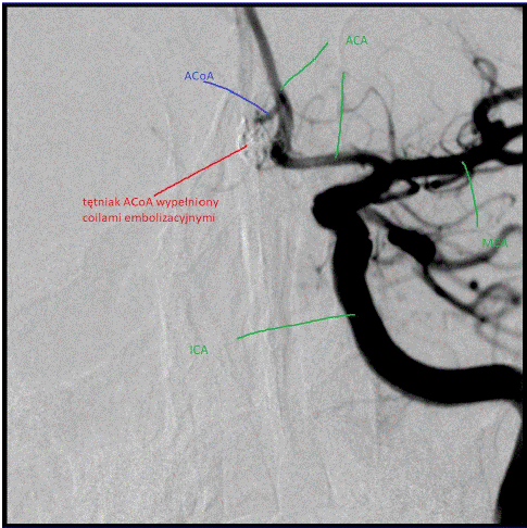
We present the case of a young man (J.J. 35 years old) who was admitted to the Cardiac Intensive Care Unit due to lateral wall myocardial infarction. His wife called for emergency medical services because he became unconscious, wheezed and vomited. The event took place during sexual intercourse. His wife, who was a nurse, put him in the recovery position, but choking could not be ruled out. The emergency medical team found him unconscious, with regular but fast heart rate and respiratory failure. He was intubated, excess secretions were removed from his airways by suctioning, and his breathing had to be helped by a ventilator. Because ECG showed lateral wall myocardial infarction (Figure 1a & 1b), the patient was transferred directly to the catheterization laboratory. A stomach tube was inserted and ASA 300 mg and clopidogrel 600 mg were administered. Also, 5000 U of heparin were used. Coronary angiography showed no stenotic lesions in the coronary arteries. The first measurement of high-sensitivity troponin I levels (hsTnI) was 1,300ng/L (normal value <35 ng/L). The second measurement, taken 3 hours later, was 13,000 ng/L. ECHO examination revealed akinesia of several segments of the left ventricle and significantly decreased Left Ventricular Ejection Fraction (LVEF), which was at 35%. Myocardial infarction was therefore determined. The patient was sedated using propofol because he was anxious and there were concerns about his intubation. The ECG performed just after coronary angiography revealed resolution of earlier changes (Figure 2). The next step was computed tomography of the head which showed an extensive subarachnoid hematoma (Figure 3). CT angiography revealed a ruptured aneurysm in the ACA region (Figure 4). Neurosurgical operation was risky because the patient received heparin and two antiplatelet drugs. A decision was made to embolize the aneurysm percutaneously. The patient was hemodynamically stable. The percutaneous embolization of the aneurysm was successfully performed. The patient was subsequently admitted to the Intensive Care Unit. However, in spite of all of the above mentioned interventions, his status worsened, as brain swelling increased. On the second day after admission, brain death was diagnosed and the patient was declared dead.

Figure 1a: The first ECG (limbal leads) – ST-segment elevation in leads I and aVL.

Figure 1b: The first ECG (precordial leads) – ST-segemnt elevation in lead C6.

Figure 2: The ECG after coronary angiography shows the resolution of ST-segment elevation.

Figure 3a and 3b: Computed tomography image - green lines indicate the anterior cerebral arteries; red line indicate an aneurysm of the artery communicans anterior.

Figure 4a: Computed tomography - red lines show subarachnoid hemorrhage.

Figure 4b: Computed tomography – red lines indicate bleeding into lateral ventricles of the brain.

Figure 5a: Angiography: An aneurysm of the artery communicans anterior before treatment; ACA: Anterior Cerebral Artery; MCA: Middle Cerebral Artery; ICA: Internal Carotid Artery.

Figure 5b: Angiography: An aneurysm of the artery communicans anterior (ACoA) after treatment (coils filling the aneurysm); ACA: Anterior Cerebral Artery; MCA: Middle Cerebral Artery; ICA: Internal Carotid Artery.
Commentary
We present this case to remind both cardiologists and neurologists about the relation between the brain and the heart. Different electrocardiographic abnormalities can be found in patients with cerebrovascular events [1,2,3]. ECG changes can cause difficulties in diagnosing and making therapeutic decisions regarding patients with a stroke. ECG changes are found in 30-50% of patients with a stroke and without concomitant cardiovascular diseases. These changes include a wide spectrum of abnormalities such as arrhythmias, prolonged QT segment, or ischemia-like changes (ST-segment depression, ST-segment elevation, negative T-wave). In the case of subarachnoid hemorrhage, ischemia-like changes in ECG are especially common (they are found in 50-100% of patients) [2,3,4]. Those changes can be transient and disappear without inducing myocardial necrosis, but in some cases an excessive sympathetic activity can lead to myocardial necrosis directly or by inducing vasospasm. The origin of ECG abnormalities in patients with a stroke is not yet established. One hypothesis suggests that they result from an imbalance between two parts of the autonomous nervous system. Usually, there is a prevalence of sympathetic activation [2,3]. Excessive sympathetic activation can lead to a structural damage of the heart or to arrhythmias. Since hypomagnesemia has been observed in SAH, another hypothesis suggests that it can facilitate vasospasm of both the cerebral and coronary arteries. In the presented case, it is likely that myocardial ischemia followed by myocardial necrosis was provoked by a redistribution of blood flow due to vasospasm of the intramuscular coronary arteries (myocardial infarction type 2). We conclude that, in the case of this patient, the first symptom being the loss of consciousness (in a patient who had no arrhythmias and no conduction disorders in the heart) suggested a cerebrovascular event rather than a heart attack. But on the other hand, ECG results were so suggestive of the latter that the right choice of a primary medical intervention was very difficult.
References
- Grabczewska Z, Swiatkiewicz I, Kubica J. ECG changes in patients with stroke. Kardiol Pol. 2009; 67: 196-9.
- Togha M, Sharifpour A, Ashraf H, Moghadam M, Sahraian MA. Electrocardiographic abnormalities in acute cerebrovascular events in patients with/without cardiovascular disease. Ann Indian Acad Neurol. 2013; 16: 66-71.
- Katsanos AH, Korantzopoulos P, Tsivgoulis G, Kyritsis AP, Kosmidou M, Giannopoulos S. Electrographic anbormalities and cardiac arrhythmias in structural brain lesion. Int J Cardiol. 2013; 167: 328-34.
- Koochaki E, Mohamadali A, Majid M, Alizargar J. Electrocardiograph changes in acute ischemic cerebral stroke. J Appl Res. 2012; 12: 53-8.