Case Report
Austin J Clin Case Rep. 2024; 11(1): 1312.
First Report of a Colopancreatic Fistula in Crohn’s Disease
Margies R¹*; Oberholzer K²; Seidmann L³; Bouzakri N¹; Mann C¹; Lang H¹; Horisberger K¹
¹Department of General, Visceral and Transplant Surgery, University Medical Center of the Johannes-Gutenberg-University, Mainz, Germany
²Department of Radiology, University Medical Center of the Johannes-Gutenberg-University, Mainz, Germany
³Department of Pathology, University Medical Center of the Johannes-Gutenberg-University, Mainz, Germany
*Corresponding author: Margies R Department of General, Visceral and Transplant Surgery, University Medical Center of the Johannes-Gutenberg-University, LangenbeckstraΒe 1, 55131 Mainz, Germany. Tel: 00491633024260 Email: rabea.margies@unimedizin-mainz.de
Received: November 24, 2023 Accepted: December 30, 2023 Published: January 05, 2024
Abstract
Penetrating complications in the sense of a fistula is a frequent problem in Crohn’s Disease; however, fistula formation into the tail of the pancreas has never been described. We report the case of a colopancreatic fistula in Crohn’s Disease, which has been successfully resected.
Keywords: Crohn’s disease; Colopancreatic fistula; IBD surgery
Abbreviations: CD: Crohn’s Disease
Introduction
Penetrating complication in Crohn's Disease in the sense of a fistula occurs in about 35% of patients with Crohn's disease. A recent population-based database analysis estimated the cumulative incidence of fistulizing CD at 20 years from diagnosis to be 50%, of which 31% were internal abdominal fistulae [1]. While some of them are rather common (entero-sigmoid or entero-entero fistulae), others are rarely encountered (e.g. entero- or colon-duodenal) [2].
Here, we report a case of a patient with a colopancreatic fistula in Crohn's colitis mimicking a cancer of the left flexure. To the best of our knowledge, there is no existing written evidence describing a histopathologically proven colo-pancreatic fistula.
Case Report
A 51-year-old woman presented to our emergency department with abdominal pain and the clinical picture of a subileus. She had a history of Crohn's Disease since 1999. In 2004, a right hemicolectomy was performed for perforation of the ascending colon. In 2015, she underwent a resection of the anastomotic region due to a stenosis of the ileo-ascending anastomosis. Reconstruction had to be performed as an end-to-end ileo-transverse anastomosis. In addition, a stenosis of the anal canal was intermittently dilated, most recently in 2021. Medical treatment was started in July 2020 with Azathioprin and in October 2021 with Infliximab.
On presentation in October 2022, the patient reported symptoms of intermittent nausea and diffuse spasmodic abdominal pain of several weeks' duration. Clinical examination revealed a distended abdomen with mild tenderness without muscular defense. The laboratory results showed an elevated CRP of 175mg/l and normal white blood cell count (4,4/nl). The last colonoscopy, performed in 2021, had already revealed a stenosis in the anastomosis area, which couldn´t be passed with the endoscope.
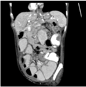
CT scan showed a stenosis in the region of the ileotransverse anastomosis with surrounding inflammatory reaction and suspicion of an abscess extending to the tail of the pancreas (Figure 1). A malignant process could not be ruled out in differential diagnostics.

Figure 1: Coronal CT reconstruction showing the connection between colon and the pancreas parenchyma.
We started to treat the patient in hospital with antibiotics and a nasogastric tube. To complete the diagnostic workup, we performed a hydro-MRI, which didn’t show any other stenosis than the already known in the ileotransverse region but reinforced the suspicion of a malignancy. Laparotomy was indicated to resect the anastomotic region.
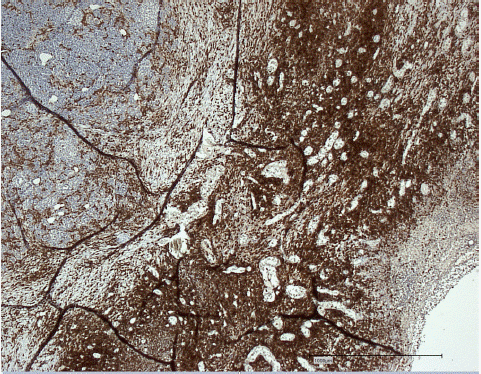
During surgery, the left transverse colon was found to be bulky and severely stenotic with infiltration into the tail of the pancreas. We performed an oncological extended left hemicolectomy with en-bloc resection of the tail of pancreas. The reconstruction was carried out as a side-to-side isoperistaltic ileosigmoid anastomosis and a drain was placed. The histopathological examination revealed a pronounced chronic florid, transmural, fistulizing and abscessing inflammation showing evidence of a fistula into the pancreatic parenchyma. No signs of malignancy were found (Figure 2).

Figure 2: Fistulizing inflammation with proliferation of CD45-positive lymphocytes (right) involving the CD45-negative pancreatic parenchyma (left). Immunostaining CD45
The patient had an uneventful postoperative course and was discharged on the ninth day in good condition with no postoperative complications, in particular no pancreatic fistula. Four-week follow-up showed normal postoperative findings with no further complications.
Discussion
Crohn's Disease is characterized by transmural inflammation, which predisposes patients to the formation of fistulae to adjacent organs as a typical complication [3]. Intestinal fistulae can occur anywhere in the gastrointestinal tract and can be associated with significant morbidity and impaired quality of life. Fistulae of colonic origin are very rare and account for about 5% of all intestinal fistulae, their typical origin is orally from an inflammatory stenosing segment [4].
A stenosis as the origin of the fistula often makes it impossible to perform a complete colonoscopy. Due to the concomitant risk of malignancy, surgical resections should be performed oncologically. In addition, in our case, preoperative imaging explicitly suggested malignancy. This could not be excluded until the final histopathological analysis.
One single case report describes a patient with a gastrocolic fistula, which ended at the surface of the pancreas. In this case, no pancreatic resection had to be performed [5]. To the best of our knowledge, a fistula between the colon and the pancreas parenchyma in Crohn's Disease has not been described yet in the current literature.
Conclusion
This is the first description of a colopancreatic fistula in penetrating Crohn's Disease. Left hemicolectomy en-bloc with the tail of the pancreas allowed removal of the entire fistula.
Author Statements
Conflict of Interest
The authors report no conflict of interest.
Author Contributions
RM wrote the short report. KO provided CT and MRI imaging. LS provided histological graphics. KH and HL supervised the project. All contributors have approved the final manuscript.
Funding
No specific funding has been received.
Data Availability Statement
All data or information generated during this case report is included in this published article.
References
- Schwartz DA, Tagarro I, Carmen Díez M, Sandborn WJ. Prevalence of fistulizing Crohn’s disease in the United States: estimate from a systematic literature review attempt and population-based database analysis. Inflam Bowel Dis. 2019; 25: 1773-9.
- Myrelid P, Soop M, George BD. Surgical planning in penetrating abdominal Crohn’s disease. Front Surg. 2022; 9: 867830.
- Cosnes J, Gower-Rousseau C, Seksik P, Cortot A. Epidemiology and natural history of inflammatory bowel diseases. Gastroenterology. 2011; 140: 1785-94.
- Michelassi F, Stella M, Balestracci T, Giuliante F, Marogna P, Block GE. Incidence, diagnosis, and treatment of enteric and colorectal fistulae in patients with Crohn’s disease. Ann Surg. 1993; 218: 660-6.
- Booth AT, Forster E, Curran T, George V. Gastrocolic fistula involving the pancreas in a patient with Crohn’s disease. Am J Gastroenterol. 2019; 114: 1823-4.