
Case Report
Austin J Clin Case Rep. 2024; 11(1): 1315.
Undifferentiated Sarcoma Crushed Intraoperatory Analysis: A Case Report
Eliezer Villanueva, MD; Alma Ortiz Plata, PhD; Martha Lilia Tena-Suck, MD*
1Servicio de Neurocirugía, Instituto Nacional de Neurología y Neurocirugía, Ciudad de México
2Laboratorio de Neuropatología experimental. Instituto Nacional de Neurología y Neurocirugía. Ciudad de México
3Departamento de Neuropatología. Instituto Nacional de Neurología y Neurocirugía. Ciudad de México
*Corresponding author: Martha Lilia Tena-Suck, MD Department of Neuropathology. National Institute of Neurology and Neurosurgery, México City, Av. Insurgentes sur no 3877, colonia la joya, Delegación Tlalpan, Cuidad de México, Mexico Email: mltenasuck@gmail.com
Received: February 02, 2024 Accepted: March 13, 2024 Published: March 20, 2024
Abstract
Sarcomas variety correspond to 1% of all cases of adult tumors, with 5-10% of those classified as Undifferentiated Pleomorphic Sarcomas (UPS / PUS) or fibro-Histiocytoma malignant or pleomorphic sarcoma. This is a rare tumor of the brain. The case corresponds to a 46-yo male, with dipsomania three times per week, with intention tremor in the right hand, then starts to shout and underwent right oral commissure deviation and speech arrest. She was carried to a hospital where Phenytoin 100 mg every 12 hours. Two months later she presented one similar crisis, and three months later, presented disorientation, dizziness, confusion, and strength decrease. She was hospitalized with epileptic syndrome and memory disturbance diagnosis. The magnetic resonance imaging demonstrated a left frontal tumor compatible with glioblastoma diagnosis. He underwent surgery and a trans-operative study was requested, showing pleomorphic, atypical large cells with large and prominent nucleoli and clear nuclei cells with small nucleoli. It was diagnosed as malignant pleomorphic neoplasia and was deferred for definitive study. In the definitive study the same image with abundant pleomorphic giant cells, alternating with small and spindle cells with extensive necrosis were seen, the cells were positive for vimentin, alpha1 anti-trypsin, lysozyme and fascin however, was negative for GFAP, and cytokeratin, CD34, HMB45. Making the diagnosis of this type of neoplasms is very difficult since the differential diagnoses are with cells of large, small, pleomorphic and elongated cells, with a necrotic background. The diagnosis of glioblastoma was discarded, and the options were adenocarcinoma, melanoma and sarcoma. In this case the immunohistochemistry gave us the diagnosis of Undifferentiated Pleomorphic Sarcoma (UPS).
Keywords: Undifferentiated pleomorphic sarcomas; Sarcomas; Cerebral tumors; Intraoperative squash smears.
Introduction
Sarcomas corresponds to 1% of all cases of adult malignancy, with 5-10% of those tumors classified as undifferentiated pleomorphic sarcomas (UPS/PUS) and represented 0.1-4.3% of primary intracranial sarcomas [1]. They recommended renaming the disease as "Pleomorphic Undifferentiated Sarcoma after the recent World Health Organization (WHO) classification [1]. High-grade sarcomas showing no line of differentiation [1]. The first report of the occurrence of MFH in Central Nervous System (CNS) was assumed by Gonzalez-Vitale et al in 1976 [2]. Intracranial UPS/PUS is characterized by an earlier age of onset and usually poorer prognosis compared to extracranial undifferentiated sarcomas or brain primaries tumors [1]. UPS/PUS is known to occur in previously abnormal bone affected by radiation, infection, and trauma. It is also associated with Paget's disease and fibrous dysplasia and offerings with varying degrees of bone destruction and mimics meningioma or glioma when is intraparenchymal located [2,3]. Few cases have been reported through, a crushed intraoperative analysis and its diagnosis can be much discussed and may be a challenger [4]. The aim of this word is a report of a rare case of cerebral undifferentiated pleomorphic sarcoma diagnosed by crushed intraoperative analysis.
Case Report
Forty-six years old male, with history of intense alcoholism, three times a week, starts with right hand intension tremor, ¿shout? dawn and presented deviation of the right labial commissure and speech impairment, and he went to the hospital and phenytoin 100 mgrs., every 12hrs were started. Two months later he presented a similar crisis, three months later he presented disorientation, dizziness, confusion, and decreased strength. With the diagnosis of epileptic syndrome and loss of memory, he was admitted to this hospital. MRI showed a left frontal lesion (Figure 1a and 1b). A right temporoparietal cystic lesion with ring reinforcement and perilesional edema was found and a glioblastoma was suspected. MRI showed an extensive lesion with central necrosis (Figure 1c and 1d). On physical examination, the patient was confused, delirious, drowsy, with a full disproportionate dense right pyramidal syndrome and underwent surgical resection. Right frontal- temporal craniotomy was performed and the lesion was resected, measuring 40x340 mm, soft friable white, and necrotic. Sample was sent for intraoperative analysis. A study was carried out by crushing and staining with H&E staining. The smears at low power showed a dense hypercellular smears, small, spindle a heterogenous cells or aberrant clustering of the cells and background densely necrotic was observed (Figure 2a and 2b), at high power the neoplastic cells were pleomorphic(Fig. 2c), with nuclear atypia (Figure 2d), cells of all sizes, with eosinophilic cytoplasm with gemistocytic appearance (Fig. 2e), multinucleated giant cells with large nucleoli (Figure 2e and 2f), it is striking that some cells the nucleus is clear and the one a small nucleolus, and some cells showed intracytoplasmic inclusion(Figure 2h).
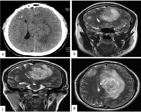
Figure 1: CT scan showing a heterogeneously enhancing hypodense infratentorial lesion in left cerebellar hemisphere and vermis compressing and displacing the fourth ventricle.
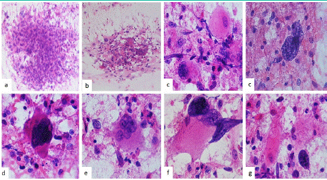
Figure 2: Intraoperative analysis. Microphotograph the smears at low power showing a population and loose aggregates of small, pleomorphic spindle cells in (a) and background densely necrotic was observed and chronic inflammation commonly accompanies this tumor in (b). (c) At high power showing small, spindle and pleomorphic neoplastic cells on them appears as finely granular background(H&Ex200). (c) Cellular atypia and hyperchromatic nuclei with multinuclear appearance with small nucleoli and range of glial processes (d), some cells with large eosinophilic cytoplasm with bizarre nuclei with multifragmentation appearance (Figure 2e). Multinucleated giant cells with large some nucleoli’s (e) and (f), showed it is striking that some cells with nucleus in different ways or different shame, in (g) those tumors nuclei can display a range of others changes, including intranuclear an intracytoplasmic inclusion or bizarre, anaplastic and pleomorphic shapes (Original magnifications H&E x400).
The cytological diagnosis was malignant pleomorphic neoplasm and deferred for definitive study. Histological sections revealed pleomorphic cell neoplasm (Figure 3a and 2b) with extensive necrosis (Figure 3c). Immunohistochemistry was positive for vimentin (Figure 3d), desmin, fascin, lysozyme (Figure 3e), p53, alpha1 anti-trypsin (Figure 3f), and higher Ki-67 li. and it was negative for S-100 protein, smooth muscle actin, epithelial membrane antigen, cytokeratin GFAP and CD34. The diagnosis of pleomorphic malignant histiocytoma or undifferentiated sarcoma was made.
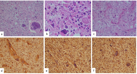
Figure 3: Microphotograph showing histopathology of the tumor, undifferentiated pleomorphic sarcoma with multinucleated giant cells (a)(H&Ex200), and (b), a close up of those giant cells, (c) that shows atypical mitotic figure and necrosis (H&E ×400 magnification). Immunohistochemistry was positive for vimentin (d), lysozyme (e), and alpha1 anti-trypsin in (f) (original magnifications, IHQ stain x400).
Discussion
Radiologically MFH/PUS is seen as large lobulated mass with hypo to isointense regions and has heterogeneous enhancement on giving contrast. Hypointense regions are indicative of necrosis. Typical radiologic findings including purely osteolytic, aggressive, destructive tumor growth with frequent soft tissue involvement, and periosteal reactions. Radiological differential diagnosis of MFH/PUS includes meningioma, osteosarcoma, chondrosarcoma, osteochondroma, dedifferentiated chordoma, plasmacytoma, lymphoma, and metastasis [5].
Histologically this is a tumor formed by pleomorphic cells and sometimes giant cells with multiple hyperchromatic irregular and bizarre nuclei and spindle cells may be seen. Histologically involves of atypical fibroblasts and histiocytes in variable sizes and proportions [1]. The diagnosis of MFH/PUS requires exclusion of another dedifferentiated pleomorphic sarcoma [1]. Four histological subtypes of MFH/PUS have been described [1]. They are storiform-pleomorphic, myxoid, giant cell and inflammatory subtype. The immunohistochemical approach reveled positive immunoreaction for vimentin, alpha-1-antitrypsin, lysosomes, fascin, and p53, and negative for SMA, S100 protein, desmin, GFAP, cytokeratin, and MyoD1. Those tumors are common alterations of certain genes, including RB1, PTEN, and DKK1 (a WNT pathway inhibitor gene) [1]. The cytopathological diagnostic is not easy to do and is very controversial. The cells with marked pleomorphism with multinucleated giant cells, with bizarre nuclei and abundant cytoplasm are thought to be characteristic cytologic findings for identifying the histiocytic features of this tumor with gemistocytic appearance [4]. The differential diagnosis includes, all the atypical and pleomorphic sarcomas and carcinomas, anaplastic oligodendrocytes and GBM [5].
The GBM is a malignant tumor that could show by crushed study a great variety of cells, small, elongated, large, oligodendrocytes, atypical gemistocytes, giant cells, pleomorphic cells, bizarre nuclei, with large nuclei, hyperchromatic, with small nucleolus and extensive necrosis in a fibrillar background [6]. GBM commonly exhibit several histological distinct regions including the classical astrocytoma vs anaplastic type, those cells including spindle, small cells or gemistocytes, anaplastic, pleomorphic and giant cells with sausage-shaped or elongated nuclei, halos and some remain of channels of vascular proliferation in a glial matrix [5,6]. Among the differential diagnoses that we must consider in the presence of giant cells, multinuclear, or small, and spindle cells with bizarre and hyperchromatic nucleoli, in fibrillar background and necrosis debris [5,6].
Cytology analysis would be useful to rule out other small round cell brain tumors including gliomas, lymphomas, carcinomas, and germinoma. Immunohistochemical analysis including tests for CD99, calretinin, and WT1 would help to suggest CIC-rearranged USRCSs and distinguish them from Ewing sarcomas characterized by CIC gene rearrangement, most commonly CIC-DUX4 fusion [1,5,6].
Cytological diagnosis of primary spindle cell lesions extremely challenging as the differential diagnosis includes a variety of such as sarcomatoid/metaplastic carcinomas, sarcomas and melanomas [1]. The intraoperatory report should indicate that the tissue does not correlate with the neuroimaging.
Conclusion: The diagnosis of PUS is a diagnosis of exclusion, that showed a dispersed population of malignant pleomorphic, bizarre, small, spindle and few multinucleated tumor giant cells. That clinically and histologically it was confused as glioblastoma. It is really a challenge for pathologists.
Author Statements
Declaration of Conflicting Interests
The author(s) declared no potential conflicts of interest with respect to the research, authorship, and/or publication of this article.
Funding
The author(s) received no financial support for the research, authorship, and/or publication of this article.
Ethics Approval
Ethical approval to report this case was obtained from mother of the patient.
References
- Fletcher CDM, Bridge JA, Hogendoorn PCW, Mertens F. World Health Organization classification of tumours of soft tissue and bone. 4th ed. Lyon: International Agency for Research on Cancer (IARC). Brain metastasis from sarcoma is rare, thus limited information is available. 2013.
- Rivero-Garvía M, Márquez-Rivas J, Rivas E, Medina-López D, Quiroga-Cantero E. Primary cerebral radiotherapy-induced rhabdomyosarcoma: treatment with intraoperative carmustine implants. Pediatr Hematol Oncol. 2013; 30: 1-3.
- Tos AP. Classification of pleomorphic sarcomas: where are we now? Histopathology. 2006; 48: 51-62.
- Ito M, Ishikawa M, Kitajima M, Narita J, Hattori S, Endo O, et al. A case report of CIC-rearranged undifferentiated small round cell sarcoma in the cerebrum. Diagn Cytopathol. 2016; 44: 828-32.
- Sharifabadi AH, Haeri H, Zeinalizadeh M, Zargari N, Razavi AE, Shahbazi N, et al. Intraoperative consultation of central nervous system lesions. Frozen section, cytology or both? Pathology - Research and Practice 2016; 212: 179-184.
- Kobayashi K, Ando K, Ito K, Tsushima M, Morozumi M, Tanaka S, et al. Accuracy of intraoperative pathological diagnosis using frozen sections of spinal cord lesions. Clinical Neurology and Neurosurgery 2018; 167: 117-121.