Letter to Editor
Austin J Clin Case Rep. 2024; 11(4): 1330.
The Burden of Polyarticular Gout with Tophi: Severe Trauma to the Bones and Soft Tissues of the Left Foot - An Inquiry into Therapeutic Approaches
Jin Peng²; Ying Zhang²; Jiashu Xie²; Dongpeng Zhang¹*
¹Department of Orthopedics, Gansu Provincial Hospital of Traditional Chinese Medicine, PR China
²Gansu University of Chinese Medicine PR China
*Corresponding author: Dongpeng Zhang Gansu Provincial Hospital of Traditional Chinese Medicine, No. 418 Guazhou Road, Qilihe District, Lanzhou, 730050, Gansu Province, China. Email: jingqian135@qq.com
Received: July 22, 2024 Accepted: August 05, 2024 Published: August 12, 2024
Letter to Editor
Dear Editor,
Gout is a prevalent disease in China that significantly affects both the physical and mental health of patients. In recent years, the incidence of gout has risen annually, correlating with improvements in living standards and dietary changes. This trend is accompanied by an increase in the prevalence of gout stone. I During the early stages of gout, urate crystals are most likely to accumulate in the first metatarsophalangeal joint [1]. With recurrent episodes of the disease, not only do uric acid deposits in the toe joints become exacerbated, but gout stones may also develop in the knee, wrist, and other anatomical areas [2]. In the advanced stage of gout, these stones can lead to acute gouty arthritis, resulting in significant destruction of bone joints and surrounding soft tissues. This article aims to address the issue of polyarticular gouty stones, highlighting a case involving severe damage to the bones and soft tissues of the left foot. A detailed account of this case follows, with a focus on potential treatment options.
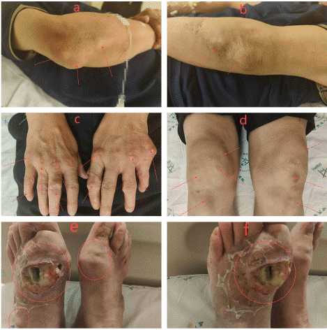
The patient under consideration is a 58-year-old male who was admitted to the hospital due to recurrent gout, accompanied by localized erythema, swelling, and rupture of the left foot. He has a history of gout spanning 19 years, with the formation of urate crystals approximately 10 years ago that have progressively increased over time. During episodes of acute gout flare, the patient has intermittently utilized colchicine and allopurinol, occasionally resorting to corticosteroid injections for symptomatic relief. The physical examination revealed the presence of urate crystals of varying sizes in multiple joints including both elbows, hands, bilateral knees, and feet. Notably, the left metatarsophalangeal joint exhibited the most pronounced accumulation, evidenced by significant local swelling, elevated skin temperature, and a notable skin rupture measuring approximately 4 cm x 4 cm (see Figure 1a-f).

Figure 1a-f: Physical examination findings: Gout stones of different sizes were seen in both elbows, hands, knees and feet, and gout stones in the left metatarsophalangeal joint were the most obvious, with local swelling, high skin temperature, and one skin rupture (size about 4cm x 4cm).
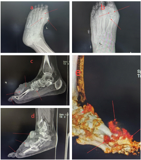
Radiographic analysis via X-ray demonstrated bone destruction localized to the left foot, with the first phalanx showing the most severe damage, in addition to marked soft tissue edema in the surrounding area (see Figure 2a-b). A Computed Tomography (CT) scan, augmented by three-dimensional reconstruction of the left foot, corroborated these findings, revealing multiple urate deposits, sustained bone loss, particularly in the first phalanx, and pronounced soft tissue swelling (Figure 2c-e). Laboratory results indicated elevated levels of uric acid (860 umol/L), a significant erythrocyte sedimentation rate (65 mm/H), an increased C-reactive protein level (66.5 mg/L), and an elevated white blood cell count (12 x 10^9/L).

Figure 2a-b: Overall survival, autologous stem cell transplant (ASCT) versus no ASCT (p=0.12).
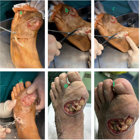
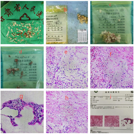
Following the patient's admission, a thorough examination of the left foot was conducted under general anaesthesia, accompanied by the excision of tophi. To facilitate the procedure, a tourniquet was applied to the left lower limb. The excision of the gouty tophi was performed using an orthopaedic spatula, while the surrounding necrotic tissue was meticulously debrided with a cold knife (Figure 3a-d). Postoperatively, the patient received continued symptomatic management, which included dressing changes, anti-infective therapy, urine alkalization, analgesia, and fluid resuscitation. The patient's recovery proceeded satisfactorily, as illustrated in Figure 3e-f. Pathological examination revealed the presence of gouty calculi in the left foot, accompanied by chronic foreign body granulomatous inflammation (refer to Figure 4a-i). Following the intervention, the patient experienced a significant improvement in symptoms, and no further necrotic tissue or gouty stones were observed in the left foot.

Figure 3a-d: The gout stone in the left foot was removed with an orthopaedic spatula and the necrotic tissue around it was removed with a cold knife (figure.3a-d). e-f: good recovery after surgery.

Figure 4a-i: Pathological examination revealed the presence of gouty tophi in the left foot, accompanied by chronic foreign body granulomatous inflammation.
Gout is frequently encountered in clinical practice; however, the occurrence of conditions resulting in significant bone destruction and deep infection remains rare. Effective management of gout necessitates an initial enhancement of dietary habits, followed by proactive control of uric acid levels [3]. In cases where gout has led to the formation of stones associated with bone and soft tissue damage, surgical intervention for stone removal and management of any resultant infections is imperative. Following surgery, a combination of pharmacological treatment—incorporating uric acid-lowering medications and traditional Chinese herbal decoctions—alongside rehabilitation therapies has been shown to produce superior clinical outcomes.
Author Statements
Conflict of Interest
The authors have no financial disclosures or other conflicts of interest to report related to the content of this article.
References
- Zhang Y, Xu SQ, Li P, Zhang WJ, Wang Y. Clinical study of staging operation for large gouty stone on the first metatarsophalangeal joint. Zhongguo Gu Shang. 2020; 33: 274-7.
- Grassi W, De Angelis R. Clinical features of gout. Reumatismo. 2012; 63: 238-45.
- Keller SF, Mandell BF. Management and Cure of Gouty Arthritis. Med Clin North Am. 2021; 105: 297-310.