Letter to Editor
Austin J Clin Case Rep. 2024; 11(4): 1331.
A 16-Year-Old Girl’s Journey to Curing Idiopathic Scoliosis: A Case Report
Guifu MA¹; Wendong Xie²; Ruixiong Nan³; Xiufen Si¹*
¹Orthopedics Department, Gansu Provincial Hospital, PR China
²Gansu University of Chinese Medicine, PR China
³The First People’s Hospital of Lanzhou City, Gansu Province, PR China
*Corresponding author: Xiufen Si, Department of Orthopedics, Gansu Provincial Hospital, No.204, Donggang WestRoad, Lanzhou, 730000, Gansu Province, PR China. Email: 527403432@qq.com
Received: July 22, 2024 Accepted: August 05, 2024 Published: August 12, 2024
Letter to Editor
Dear Editor,
Idiopathic scoliosis is a common adolescent skeletal condition characterized by an asymmetrical curvature on both sides of the spine. This condition can lead to health issues such as back pain, breathing difficulties, shoulder asymmetry, and even affect the patient’s mental health. Orthopedic spine surgery is a surgical procedure that corrects scoliosis by adjusting the curve of the spine. During the procedure, special implants, such as metal rods and pedicle pins, are used to secure and stretch the spine. This method is effective in correcting scoliosis and improving the patient's appearance and function.
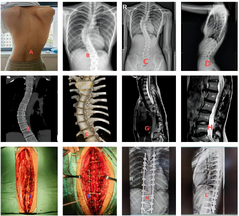
The patient, female, 16-year-old, 174 cm tall, had been hospitalized for 12 days due to dyspnoea and pain in her chest and lower back. She reported that scoliosis was detected during a physical examination at the age of 10, and on the advice of her doctor, she wore an orthosis for 1 year, which improved the degree of scoliosis compared to before. As she grew and developed, the scoliosis gradually worsened, the orthosis lost its effect, and she came to the hospital with dyspnoea, pain in the chest and lower back, and worsened symptoms while walking. A physical examination revealed an "S"-shaped scoliosis of the spine with inconsistent shoulder heights on both sides (Figure 1A). X-ray examination, CT scan and 3D reconstruction also showed "S"-shaped scoliosis (Figure 1B-F). MRI showed: scoliosis deformity, accompanied by lumbar segmental scoliosis. MRI showed: scoliosis deformity and multiple disc degeneration in the lumbar segment, but without the compression of the spinal cord (Figure 1G-H).

Figure 1: A: Scoliosis of the spine in “S” shape, with inconsistent shoulder heights on bothsides; B-F: X-ray examination, CT scan and 3D reconstruction show: scoliosis of the spine in “S” shape; G-H: MRI shows: scoliosis deformity, with disc degeneration in lumbar segments, but without the compression of the spinal cord; I: obvious scoliosis was seen during the operation; J: after the correction of scoliosis in thoracic and lumbar segments, the spinal cord was not compressed. G-H: MRI: scoliosis deformity, multiple disc degeneration in the lumbar segment, but without the compression of the spinal cord; I: obvious scoliosis was seen during the operation; J: after correcting the scoliosis of the thoracic and lumbar segments, the scoliosis was fixed with pedicle screws and metal rods, and the scoliosis was obviously improved after the completion of fixation; K-L: X-rays on the 15th day after the operation showed that the scoliosis was obviously improved compared with the previous one, without any abnormality in the internal fixation device.
After admission, spinal examination and scoliosis correction were performed under general anesthesia. Obvious scoliosis was seen after operation (Figure 1I). After the scoliosis of the thoracic and lumbar segments was corrected, the spine was fixed with pedicle screws and metal rods, and the scoliosis was seen to be significantly improved after the fixation was completed (Figure 1J). Two days after the operation, the patient felt a significant reduction in dyspnoea compared with the previous symptoms. X-ray examination was performed 15 days after surgery, showing that the scoliosis was significantly improved than before, without any abnormality in the internal fixation device (Figure 1K-L). After spinal correction, the girl was 178 cm tall. Three months after the examination, the patient had no dyspnoea, and the pain in the chest and lower back gradually disappeared. The patient continued to be followed for changes in the spine.
Wearing orthoses and surgical treatment are both options for idiopathic scoliosis. However, surgical treatment is preferred in the presence of organ dysfunction, along with rehabilitation for better results.
Author Statements
Conflict of Interest
The authors have no financial disclosures or other conflicts of interest to report related to the content of this article.
References
- Kuznia AL, Hernandez AK, Lee LU. Adolescent Idiopathic Scoliosis: Common Questions and Answers. Am Fam Physician. 2020; 101: 19-23.
- Peng Y, Wang SR, Qiu GX, Zhang JG, Zhuang QY. Research progress on the etiology and pathogenesis of adolescent idiopathic scoliosis. Chin Med J (Engl). 2020; 133: 483-493.
- Locke LL, Rhodes LN, Sheffer BW. Accelerated Protocols in Adolescent Idiopathic Scoliosis Surgery. Orthop Clin North Am. 2023; 54: 427-433.
- Walker CT, Agarwal N, Eastlack RK, Mundis GM, Alan N, Iannacone T, et al. Surgical treatment of young adults with idiopathic scoliosis. J Neurosurg Spine. 2022; 38: 84-90.