
Case Report
Austin J Clin Case Rep. 2024; 11(5): 1333.
Pedicle Screw Fixation Combined with Cemented Vertebroplasty in the Treatment of Kümmell Type 3 Fracture in Elderly Patients
Xiudong Liu; Qingjun Li; Dong Liu; Ning Li*
Department of Orthopedics, Gansu University of Chinese Medicine, PR China
*Corresponding author: Ning Li, Department of Orthopedics, Gansu University of Chinese Medicine, No.35 Dingxi East Road, Chengguan District, Lanzhou, 730000, Gansu Province, PR China. Email: 13083782939@163.com
Received: July 22, 2024 Accepted: August 07, 2024 Published: August 14, 2024
Abstract
Kümmell’s disease, first reported by German physician Herman Kümmell, is characterized as a delayed vertebral collapse following trauma [1] and represents a serious complication associated with osteoporotic vertebral compression fracture (OVCF) [2]. This condition is predominantly observed in elderly patients, who often sustain minor spinal trauma that can lead to symptoms such as such as kyphotic deformity collapse, pain, paresthesia, and limitations in functional capabilities as the disease progresses. Under the trend of aging population, the prevalence of Kümmell disease is increasing year by year. In cases of stage III Kümmell disease, the incomplete posterior wall can predispose patients to complications during treatment, particularly during procedures such as Percutaneous Vertebroplasty (PVP) or Percutaneous Kyphoplasty (PKP). These complications include cement leakage and displacement, which can exacerbate spinal cord compression [3]. Anterior decompression, while an option, is associated with an elevated risk of visceral injury and infection. Conversely, posterior total spinal surgery is not only lengthy and invasive but may also result in a prolonged rehabilitation period and increased complications. Therefore, the selection of an appropriate surgical intervention is critical in managing Kümmell’s disease effectively.
Keywords: Stage 3 Kümmell’s fracture; Severe osteoporosis; Spinal cord compression; Pedicle screw fixation; Cement augmentation; Minimally invasive surgery
Introduction
Kümmell Type 3 fractures refer to a fracture type of spinal compression fracture with kyphotic deformity. This type of fracture usually occurs in the thoracolumbar spine and is more common in the elderly, especially in patients with osteoporosis. Typical symptoms of Kümmell Type 3 fractures are pain, limited motion, and kyphotic deformity.
Case Report
An 82-year-old female patient with a past history of osteoporosis presented to the hospital following a fall at home before January. She reported persistent low back pain and discomfort, which was not relieved by oral non-steroidal anti-inflammatory drugs (NSAIDs). Pain was assessed using the Visual Analogue Scale (VAS), yielding a score of 6 points in the supine position and 8 points in the upright position. Physical examination revealed percussion pain over the spinous processes in the thoracolumbar region, muscle strength in both lower limbs graded as III, and bilateral lower limb numbness, with no defecation disorders. Three-dimensional reconstruction and X-ray imaging indicated a T12 vertebral fracture with an intravertebral fissure (Figure 1a-c). MRI confirmed a compression fracture of the T12 vertebral body with spinal cord compression (Figure 1d). Dual-Energy X-ray Absorptiometry (DEXA) demonstrated severe osteoporosis in the spine, with a T-score of -4.2 (Figure 2 and Figure 3). A comparative bone density table confirmed severe spinal osteoporosis (Figure 4). Considering that the patient's symptoms were serious and there was no absolute contraindication for surgery, a decision was made to proceed with surgical intervention following discussions with the patient and her family. The chosen approach was pedicle screw fixation combined with vertebroplasty, aimed at reducing spinal cord compression and restoring the spine's physiological curvature and stability. During the procedure, bone cement was injected into the T11, T12, and L1 vertebral bodies to restore their height as much as possible. Subsequently, pedicle screws were inserted after the cement had set, and the spine was further stabilized with metal rods to restore normal thoracic and lumbar curvature and stability (Figure 5a-d).
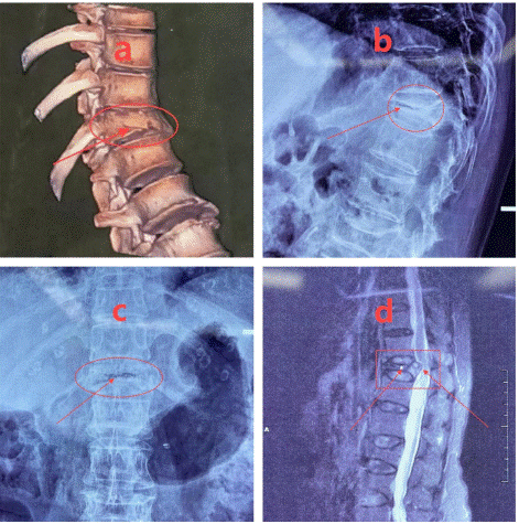
Figure 1: a-c) Three-dimensional reconstruction and X-ray showed T12 vertebral body fracture with signs of intravertebral fissure. d) The MRI scan revealed a compression fracture of the T12 vertebral body, accompanied by spinal cord compression.
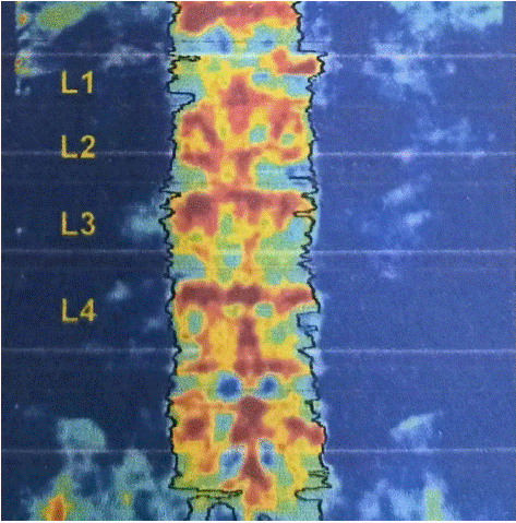
Figure 2: Dual energy X-ray bone densitometry showed that the patient had severe osteoporosis in the spine.
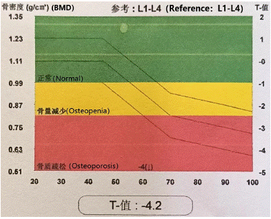
Figure 3: The T value of bone mineral density test was -4.2.
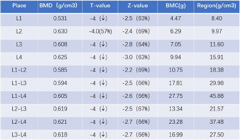
Figure 4: The bone density value comparison table showed that the patient had severe osteoporosis in the spine.
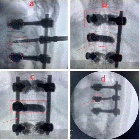
Figure 5: a-d) During the surgical procedure, bone cement was meticulously implanted into the T11, T12, and L1 vertebral bodies to achieve maximal restoration of their original height. Following the hardening of the cement, pedicle screws were subsequently inserted. The fixation was further reinforced with metal rods to restore the normal physiological curvature and stability of the thoracic and lumbar vertebrae.
Postoperatively, partial restoration of the T12 vertebral body height was achieved, with no evidence of cement leakage or migration. The patient reported significant improvement in low back pain and lower limb weakness and numbness one day after surgery. Five days post-surgery, a follow-up VAS assessment showed a marked reduction in pain scores compared to preoperative levels. The patient expressed satisfaction with the treatment and continued anti-osteoporotic medication following discharge. Ongoing follow-up of the patient's condition was planned.
Discussion
The patient presented with severe osteoporosis and spinal cord compression. Initially, the vertebral body was augmented with bone cement to maximize the restoration of vertebral height. Subsequently, pedicle screws and metal rods were utilized to reestablish the physiological curvature and stability of the spine.
This surgical approach offers several advantages:
1. It enhances the biomechanical stability of the fracture zone, facilitating the early restoration of a stable spinal structure. This intervention minimizes soft tissue strain in the lumbar region and prevents re-collapse of the injured vertebra [4].
2. It provides rapid alleviation of symptoms associated with spinal cord compression, effectively reducing the risk of surgical complications and yielding favourable clinical outcomes.
Author Statements
Conflict of Interest
The authors declare no financial disclosures or other conflicts of interest relevant to the content of this article to report.
References
- Brower AC, Downey EJ. Kümmelll disease: report of a case with se rial radiographs. Radiology. 1981; 141: 363-364.
- Zhou C, Huang S, Liao Y, Zhang F, Meng X, Tang Z, et al. Feasibility Analysis of the Bone Cement-Gelatine Sponge Composite Intravertebral Prefilling Technique for Reducing Bone Cement Leakage in Stage I and II Kümmell’s Disease: A Prospective Randomized Controlled Trial. Orthop Surg. 2023; 15: 1763-1771.
- Mo GY, Zhou TP, Guo HZ, Li YX, Tang YC, Guo DQ, et al. Long-term efficacy and safety of bone cement-augmented pedicle screw fixation for stage III Kümmell disease. Sci Rep. 2021; 11: 13647.
- Zhan Y, Bao C, Yang H, Li L, Yan L, Kong L, et al. Biomechanical analysis of a novel bone cement bridging screw system combined with percutaneous vertebroplasty for treating Kümmelll’s disease. Front Bioeng Biotechnol. 2023; 11: 1077192.