
Case Report
Austin J Clin Case Rep. 2024; 11(5): 1334.
Knee Replacement Combined with Ligament Reconstruction for Severe Knee Valgus with Knee Subluxation: A Case Report
Xiudong Liu²; Qingjun Li²; Wendong Xie²; Chao Zhang¹*
¹Department of Orthopedics, Gansu Provincial Hospital, PR China
²Gansu University of Chinese Medicine, PR China
*Corresponding author: Chao Zhang, Department of Orthopedics, Gansu Provincial Hospital, No.204, Donggang WestRoad, Lanzhou, 730000, Gansu Province, PR China. Email: zc-315@163.com
Received: July 27, 2024 Accepted: August 07, 2024 Published: August 14, 2024
Abstract
With the aging of the population and the accelerated pace of social life, knee diseases have become a major problem that seriously affects the quality of life among middle-aged and elderly people in China. Among these conditions, knee valgus with knee subluxation stands out as a severe form of knee disease, often characterized by intense pain and restricted joint mobility. The etiology of knee valgus with knee subluxation primarily stems from factors such as knee ligament damage, meniscus injury, joint inflammation. Moreover, long-term joint pathology can induce structural alterations within the knee joint, which in turn triggers knee valgus and subluxation. This condition has a serious impact on the patient’s quality of life, manifesting as pain, limited joint function, limping, etc. In severe cases, it may even lead to joint disability.
Keywords: Knee replacement; Ligament Repair; Knee valgus; Subluxation of the knee joint; Osteoarthropathy of the knee
Introduction
Knee replacement combined with ligament reconstruction is a surgical procedure that combines knee replacement with ligament reconstruction. The principle is to improve the structure and function of the knee joint through artificial joint replacement while simultaneously rebuilding the damaged ligaments to restore the stability of the knee joint.
Case Report
The patient under consideration is a 60-year-old woman who was diagnosed with osteoarthropathy of the right knee joint at a local hospital a decade ago. The diagnosis was made due to the patient's complaints of pain and limited movement in the right knee. Following the diagnosis, the patient was administered pain relief medication and acupuncture, which led to symptomatic relief and subsequent discharge from the hospital. However, over time, the patient experienced repeated episodes of knee deformity, which gradually worsened. A physical examination revealed that the patient ha right knee valgus with knee subluxation, joint space narrowing, and severe articular surface wear, as depicted in Figure 1a-d. X-ray examination further confirmed the presence of right knee degenerative disease and right knee subluxation, as shown in Figure 2a-c. MRI examination revealed that the patient was suffering from degenerative osteoarthropathy of the right knee joint, along with knee subluxation, medial and lateral meniscus prolapse, medial collateral ligament injury, bone marrow oedema, effusion in the joint cavity and suprapatellar bursa of the right knee, and swelling of the soft tissues around the knee joint, as depicted in Figure 2d-m. Upon admission, the right knee joint was examined and the lesion removed under general anesthesia. A tourniquet was concurrently applied to the right lower limb. Intraoperatively, severe osteophytes, synovial hyperplasia and ligament damage were observed in the right knee joint. The synovial hyperplasia around it was removed using an electro coagulator, while the hyperplastic bone around it was removed with a bone-biting forceps (Figure 3a-d). The proliferated bone of the medial femoral condyle was removed with a chainsaw to rebalance the joint (Figure 3e-h). Residual tissue and meniscus were excised with the electro coagulator (Figure 3i-j). Measurement of knee balance and right lower limb mechanical axis was performed along with repair of knee ligaments to ensure restoration of knee function (Figure 3k-l). Postoperative X-ray examination confirmed proper positioning of the artificial joint and ligament repair devices, with favorable joint spacing in the right knee following artificial joint replacement (Figure 4a-b). Postoperative physical examination indicated complete resolution of the right knee deformity (Figure 4c). Following knee joint replacement combined with ligament reconstruction, the patient experienced significant reduction in knee pain, improvement in joint function, and notable enhancement in quality of life.
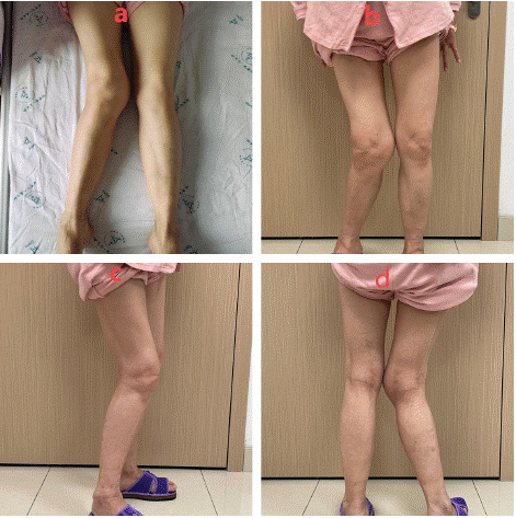
Figure 1: a-d) Physical examination showed right knee valgus with knee subluxation, narrow joint space, and severe articular surface wear.wear.
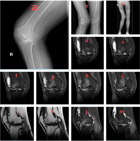
Figure 2: a-c) X-rays showed degenerative lesions of the right knee joint and subluxation of the right knee. d-m) MRI examination revealed that the patient was suffering from degenerative osteoarthropathy of the right knee joint, along with knee subluxation, medial and lateral meniscus prolapse, medial collateral ligament injury, bone marrow oedema, effusion in the joint cavity and suprapatellar bursa of the right knee, and swelling of the soft tissues around the knee joint.
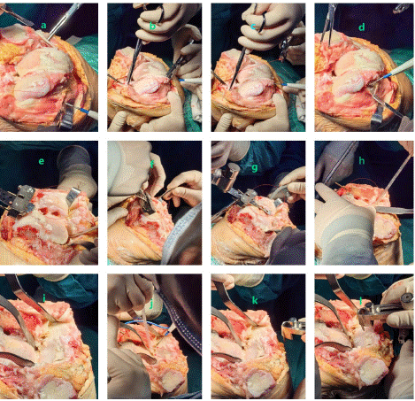
Figure 3: a-d) The synovial hyperplasia surrounding it was removed using an electrocoagulation knife, while the bony hyperplasia surrounding it was removed with a bone-biting forceps. e-h): The proliferated bone of the medial femoral condyle was removed with a chainsaw to rebalance the joint. i-j) Residual tissue and meniscus were excised with an electrocoagulation knife. k-l) Measurement of knee balance and right lower extremity mechanical axis was performed along with repair of knee ligaments to ensure restoration of knee function.
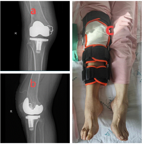
Figure 4: a-b) Postoperative X-rays examination confirmed proper positioning of the artificial joint and ligament repair devices, with favorable joint spacing in the right knee following artificial joint replacement. c) Postoperative physical examination indicated complete resolution of the right knee deformity.
Discussion
Knee replacement combined with ligament reconstruction represents the primary intervention for the treatment of severe knee valgus with knee subluxation. Postoperatively, the patient received comprehensive care aimed at preventing infection and managing pain. Additionally, the patient was instructed to engaged in structured rehabilitation exercises in order to promote the restoration of joint functionality.
Author Statements
Conflict of Interest
The authors have no financial disclosures or other conflicts of interest to report related to the content of this article.
References
- Lal H, Sabharwal VK, Tanwar Y. Total knee replacement in triple deformity with posterior subluxation of the knee joint. J Clin Orthop Trauma. 2015; 6: 113-9.
- Li T, Liu Y, Li C, Zhang H. SMOC approach for total knee arthroplasty in valgus knees. J Orthop Surg Res. 2022; 17: 120.
- Li H, Xie JW, Ding ZC, Yuan MC, Lai YH, Zhou ZK. Evaluation of the rate of post-operative dislocation in patients with ipsilateral valgus knee deformity after primary total hip arthroplasty. Int Orthop. 2022; 46: 1507-1514.
- Sun JY, Ma HY, Shen JM, Du YQ, Dong Y, Zhang YC, et al. Factors influencing knee valgus alignment in Crowe type IV hip dysplasia after total hip arthroplasty. J Orthop Traumatol. 2021; 22: 41.