
Case Report
Austin J Clin Case Rep. 2024; 11(5): 1335.
Tuberculosis of the Right Pubic Bone Complicated by Abscess of The Right Thin Femoral Muscle: A Case Report
Bo Liu1; Wendong Xie2; Weiwang Tuo2; Jin Peng2; Guifu Ma1*
¹Department of Orthopedics, Gansu Provincial Hospital, PR China
²Gansu University of Chinese Medicine, PR China
*Corresponding author: Guifu Ma, Department of Orthopedics, Gansu Provincial Hospital, No.204, Donggang West Road, Lanzhou, 730000, Gansu Province, PR China. Email: 15693318899@163.com
Received: July 27, 2024 Accepted: August 08, 2024 Published: August 15, 2024
Abstract
Tuberculosis of the pubic bone is a kind of bone tuberculosis, because the pubic bone is located at the bottom of the pelvis, and its surrounding tissues are rich in blood vessels and nerves, therefore, tuberculosis of the pubic bone is easy to invade the surrounding muscles and fascia, and cause inflammatory reaction. The reason of pubic tuberculosis complicating abscess is mainly caused by the infection of tubercle bacillus. After Mycobacterium tuberculosis invades the pubic bone, due to the poor blood supply of the pubic bone, the bacteria are easy to multiply locally, triggering an inflammatory reaction. As the inflammation intensifies, the local tissues become necrotic and liquefied, forming an abscess. After abscess formation, it will not only lead to increased pain, but may also invade the surrounding nerves and blood vessels, triggering more serious complications.
Keywords: Pubic bone tuberculosis; Femoral thin muscle abscess; Surgical treatment; Anti-tuberculosis drugs; Continuous rinsing with iodophor solution and hydrogen peroxide solution
Introduction
Tuberculosis of the pubic symphysis is a condition characterized by the presence of tuberculosis infection in the pubic symphysis, which is the joint located at the front of the pelvis where the two pubic bones meet. Tuberculosis is a worldwide public health crisis. The osteoarticular form is uncommon: it represents 2-5 % of all tuberculosis cases [1,2] and 11-15% of extrapulmonary tuberculosis. The main localization remains vertebral tuberculosis, which accounts for half of the cases [3,4]. Pubic localization is particularly rare.
Case Report
The patient was a 69-year-old woman. She reported right-sided lower abdominal pain that started six months ago and gradually worsened, with discomfort in walking, sitting and standing. Over the past month, the pain had worsened and was accompanied by low-grade fever and malaise. Physical examination showed a bulging mass in the right medial thigh, with obvious tenderness in the right pubic bone area as well as in the medial thigh (Figure 1a-c). B-mode ultrasonography showed an irregular hypoechoic mass from the right thigh to the pubic bone, which was considered to be a tuberculosis (cold abscess formation) (Figure 2). X-ray orthopedic position of the pelvis showed an uneven density in the right pubic bone (Figure 3). ECT bone scan showed an abnormally metabolically active focus of bone metabolism in the right pubic bone, and tuberculosis infection was considered in conjunction with the medical history (Figure 4).Three-dimensional reconstruction and CT scan showed: right pubic bone destruction, right pubococcygeus muscle, closed hole external muscle swelling, thin femoral muscle abscess formation (Figure 5a-d). MRI showed: right thin femoral muscle peripheral mass shadow and oozing, right pubic bone abnormal signal, combined with the history of the possibility of tuberculosis infection (Figure 6a-c) After admission, the right pubic bone and right medial thigh were examined and the lesion was resected under general anesthesia. Intraoperatively, bone destruction of the right pubic bone and abscess formation in the thin femoral and medial thigh muscle space were seen. Necrotic tissue and synovial proliferation around it were removed with a condenser knife, and the necrotic pubic bone was removed with a biting forceps. A drain was placed at the location of the lesion, and iodophor solution and hydrogen peroxide solution were continuously flushed (Figure 7a-b). Pathological examination showed: chronic granulomatous inflammation with visible caseous necrosis, tuberculosis was considered (Figure 8). Mycobacterium tuberculosis DNA test: positive for TB-DNA(+) (Figure 9). Regular oral anti-tuberculosis drugs: rifampicin, isoniazid, ethambutol pyrazinamide, and regular rechecks of liver function and renal function. After 1 year of examination, no new bone destruction or abscess formation was found.
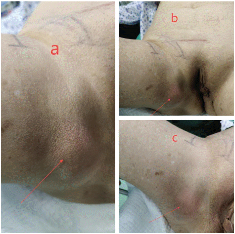
Figure 1: a-c) Physical examination showed a bulging mass in the right medial thigh, with obvious tenderness in the right pubic bone area as well as in the medial thigh.
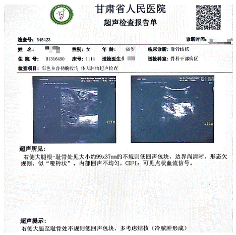
Figure 2: B-mode ultrasonography showed an irregular hypoechoic mass from the right thigh to the pubic bone, which was considered to be a tuberculosis (cold abscess formation).
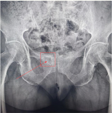
Figure 3: X-ray orthopedic position of the pelvis showed an uneven density in the right pubicbone.
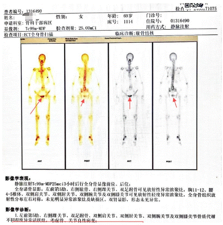
Figure 4: ECT bone scan showed an abnormally metabolically active focus of bone metabolism in the right pubic bone, and tuberculosis infection was considered in conjunction with the medical history.
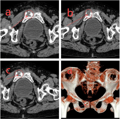
Figure 5: a-d) Three-dimensional reconstruction and CT scan showed: right pubic bone destruction, right pubococcygeus muscle, closed hole external muscle swelling, thin femoral muscle abscess formation.
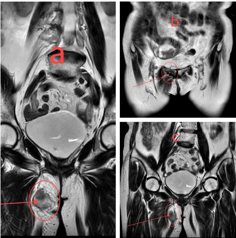
Figure 6: a-c) MRI showed: right thin femoral muscle peripheral mass shadow and oozing, right pubic bone abnormal signal, combined with the history of the possibility of tuberculosis infection.
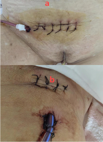
    Figure 7: a-b) A drain was placed at the location of the lesion, and iodophor solution and hydrogen peroxide solution were
continuously flushed.
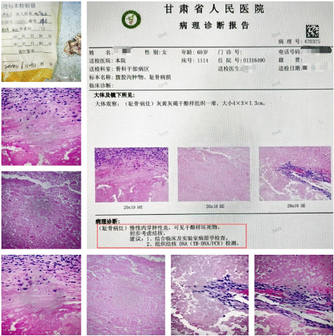
Figure 8: Pathological examination showed: chronic granulomatous inflammation with visible caseous necrosis, tuberculosis was considered.
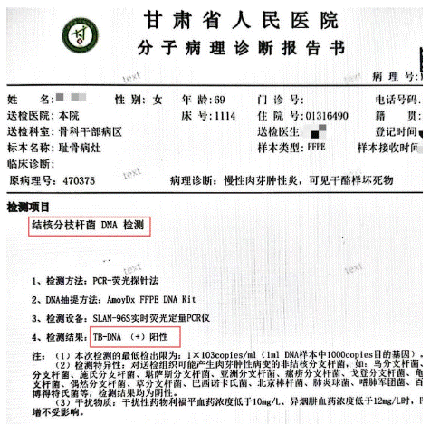
Figure 9: Mycobacterium tuberculosis DNA test: positive for TB-DNA(+).
Discussion
Drug therapy is the first choice for the treatment of pubic bone tuberculosis. Surgical treatment is needed when the following conditions occur: abscess formation bone destruction spread of the lesion ineffective drug treatment. On the basis of drug treatment and surgery, patients need to carry out rehabilitation exercises to restore muscle strength and joint mobility.
Author Statements
Conflict of Interest
The authors have no financial disclosures or other conflicts of interest to report related to the content of this article.
References
- Ellis ME, el-Ramahi KM, al-Dalaan AN. Tuberculosis of peripheral joints: a dilemma in diagnosis. Tuber Lung Dis. 1993; 74: 399-404.
- Monach PA, Daily JP, Rodriguez-Herrera G, Solomon DH. Tuberculous osteomyelitis presenting as shoulder pain. J Rheumatol. 2003; 30: 851-6.
- Benbouazza K, Allali F, Bezza A, el Hassani S, el Maghraoui A, Lazrak N, Hassouni F, Hajjaj-Hassouni N. L’ostéoarthrite pubienne tuberculeuse. A propos de deux cas [Pubic tuberculous osteo-arthritis. Apropos of 2 cases]. Rev Chir Orthop Reparatrice Appar Mot. 1997; 83: 670-2.
- Meena S, Gangary SK. Tuberculosis of symphysis pubis: A case report. J Res Med Sci. 2015; 20: 100-2.