
Case Report
Austin J Clin Case Rep. 2024; 11(5): 1336.
Lumbar Isthmic Fissure Causing Lumbar Spondylolisthesis of II Degree, Lumbar Discherniation, Lumbar Spinal Stenosis: A Case Report
Haodong Yang²; Xingwen Xie¹; Hao Liu²; Wendong Xie²; Ning Li¹*
¹Department of Orthopedics, Affiliated Hospital of Gansu University of Chinese Medicine, PR China
²Gansu University of Chinese Medicine, PR China
*Corresponding author: Ning Li, Department of Orthopedics, Affiliated Hospital of Gansu University of Chinese Medicine, No.732 Jiayuguan West Road, Chengguan District, Lanzhou,730000, PR China. Email: 13083782939@163.com
Received: July 30, 2024 Accepted: August 08, 2024 Published: August 15, 2024
Abstract
Lumbar isthmic cleft, also known as isthmic cleft of the vertebral arch or isthmic discontinuity, is a discontinuous bony defect in the isthmus between the upper and lower articular processes of the lumbar vertebral arch. Lumbar isthmic fracture may lead to serious consequences such as lumbar spine instability, slippage and disc herniation, which will bring great trouble to the patient’s life and work. First of all, how does lumbar isthmic fracture cause lumbar spondylolisthesis? A lumbar isthmic fracture weakens the connection between the lumbar vertebrae, which can cause the vertebrae to move back and forth, leading to lumbar spondylolisthesis. Lumbar spondylolisthesis is the back-and-forth movement of the upper vertebrae of the lumbar spine in relation to the lower vertebrae, which can compress the nerve roots and lead to symptoms such as pain and numbness. Secondly, lumbar isthmic fracture can also lead to disc herniation.
Keywords: Lumbar isthmic cleft; Lumbar spondylolisthesis; Lumbar disc herniation; Lumbar spinal stenosis; Surgical treatment
Introduction
A lumbar isthmic fracture affects the stability of the lumbar spine, which can increase the pressure on the discs, leading to a herniated disc, which can compress the nerve roots or spinal cord, causing symptoms such as pain, numbness, and fatigue. Finally, a lumbar isthmic fracture can lead to spinal stenosis. A lumbar isthmic fracture weakens the connecting parts of the lumbar vertebrae, which can cause the vertebrae to move back and forth, which reduces the space in the spinal canal, compressing the spinal cord and causing spinal stenosis. Spinal stenosis is a condition in which the space in the spinal canal becomes smaller and compresses the spinal cord, resulting in symptoms such as pain, numbness, and weakness.
Case Report
The patient was a 56-year-old male who spent 19 days in the hospital due to lumbar pain with numbness and weakness in both lower extremities. He reported that he started to have pain in his lower back 5 years ago, which was relieved by oral painkillers with recurrent episodes, and recently the pain in his lower back had worsened, with numbness in both lower limbs, weakness, and difficulty in walking. Physical examination showed obvious pressure and pain in the lumbar region, and the straight leg raising test was positive. x-ray examination showed lumbar 5 vertebral body degree slip (Figure 1a-b), CT scan showed lumbar 5 vertebral body degree slip, lumbar isthmic fissure, and vertebral body margins "bird's beak sign" (Figure 1c-g), MRI showed lumbar 5 vertebral body degree slip, disc herniation, and vertebral body edge "bird's beak sign" (Fig.1c-g), and MRI showed lumbar 5 vertebral body degree slip, disc herniation, and vertebral body edge "bird's beak sign". MRI showed: lumbar 5 vertebral body II-degree slip, disc herniation, spinal canal stenosis (Figure 1h-j).

Figure 1: a-b) X-rays show: lumbar 5 vertebral body II degree slip. c-g: CT scans show: lumbar 5 vertebral body II degree slip, lumbar isthmic fissure, and vertebral body margin “bird’s beak sign”. h-j: MRIs show: lumbar 5 vertebral body II degree slip, intervertebral disc protrusion, and spinal canal stenosis.
After admission, lumbar 4 vertebrae, lumbar 5 vertebrae, sacral 1 vertebra were examined and lesions were removed under general anesthesia. Intraoperatively, lumbar5 vertebral body slip, disc herniation, and spinal stenosis were seen. The proliferated bone was removed with a bone biting forceps and the disc was removed with a reamer (Figure 2a-g). The vertebral body was fixed with metal rods and pedicle pins, and the slipped vertebra was reset by slow traction (Figure 2h-l). One day after the operation, the patient's symptoms of lumbar pain, numbness of both lower limbs, and fatigue were significantly reduced. Postoperative X-ray examination showed that the lumbar vertebral slip was completely repositioned and the internal fixation device was in good position (Figure 3a-c). Follow-up of the patient's condition changes was continued.
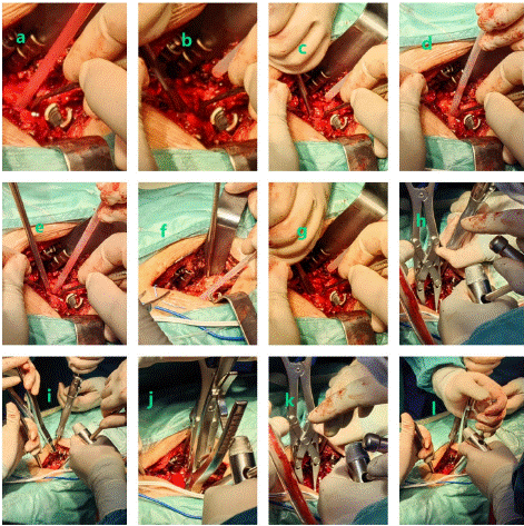
Figure 2: a-g) removal of hyperplastic bone with a bone-biting forceps and removal of the disc with a reamer. h-l: fixation of the vertebral body with metal rods and pedicle pins, and slow traction to reset the slipped vertebral body.
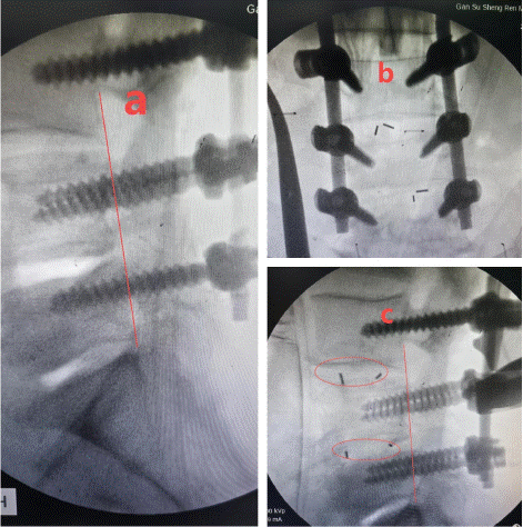
Figure 3: a-c) Postoperative X-rays show complete repositioning of the lumbar spondylolisthesis and good position of the internal fixation device.
Discussion
For patients with mild symptoms, conservative treatment is the first choice. It includes: avoiding strenuous exercise and heavy physical labour, and giving sufficient rest to the lower back. Medication: apply non-steroidal anti-inflammatory drugs, blood circulation and blood stasis activating drugs to relieve pain. Physiotherapy: such as hot compresses, massage, acupuncture, etc., can help relieve symptoms. Lumbar back muscle exercise: strengthen the lumbar back muscle strength and improve the stability of lumbar spine. For patients with severe symptoms and ineffective conservative treatment, surgical treatment is necessary. Surgical methods include: isthmus repair: surgically repairing the broken isthmus to restore lumbar spine stability. Vertebral fusion: Fusing adjacent vertebrae to eliminate slippage. Internal Fixation: Fixation of the broken isthmus using a metal internal fixator to promote healing.
Author Statements
Conflict of Interest
The authors have no financial disclosures or other conflicts of interest to report related to the content of this article.
References
- Peng JC, Guo HZ, Zhan CG, Huang HS, Ma YH, Zhang SC, et al. Comparison of pedicle screw fixation with or without cement augmentation for treating single-segment isthmic spondylolisthesis in the osteoporotic spine. Sci Rep. 2023; 13: 827.
- Raffa SJ, Luther E, Levi AD. Repair of isthmic pars interarticularis fractures: a literature review of conventional and minimally invasive techniques. J Neurosurg Sci. 2019; 63: 318-329.
- Minamide A, Akamaru T, Yoon ST, Tamaki T, Rhee JM, Hutton WC. Transdiscal L5-S1 screws for the fixation of isthmic spondylolisthesis: a biomechanical evaluation. J Spinal Disord Tech. 2003; 16: 144-9.
- Carragee EJ. Single-level posterolateral arthrodesis, with or without posterior decompression, for the treatment of isthmic spondylolisthesis in adults. A prospective, randomized study. J Bone Joint Surg Am. 1997; 79: 1175-80.