Case Series
Austin J Clin Case Rep. 2024; 11(7): 1344.
A Case Study of Two Benign Retro Rectal Tumors
El Mouhib S*; Dabbagh M; Ed-Dahby O; Bouzroud M; El Kaoui H; Bouchentouf SM; Moujahid M
Department of General Surgery, Mohammed V Military Hospital, Rabat Morocco
*Corresponding author: El Mouhib S, Department of General Surgery, Mohammed V Military Hospital, Rabat, Morocco. Tel: +2120622391529 Email: safaelmo28@gmail.com
Received: November 19, 2024; Accepted: November 27, 2024; Published: December 04, 2024
Abstract
Retrorectal tumors are a rare entity that present with unspecific clinical presentation and are often asymptomatic. They present a diagnostic and therapeutic challenge due to their varied histology and anatomical location. A pelvic MRI is recommended to evaluate the extent of the resection and determine the surgical approach. This report presents two cases of benign retrorectal tumors, a ganglioneuroma and a fibromatosis, in patients aged 69 and 25, respectively. These cases highlight the age-related variation in the presentation of retrorectal lesions and emphasize the rarity of certain benign tumors, particularly ganglioneuroma and fibromatosis, in the retrorectal space.
Keywords: Retrorectal; Presacral; Tumor; Ganglioneuroma; Fibromatosis; MRI
Introduction
Retrorectal neoplasms develop in the anatomical region between the rectum and sacrum, and they are uncommon lesions that can be difficult to diagnose due to their nonspecific clinical manifestations. The retrorectal space comprises an array of tissue types, including adipose, connective, and neurovascular components, contributing to the heterogeneity of retrorectal tumors that range from benign cysts to complex malignant masses that can invade the surrounding pelvic structures. The predominant neoplasms in this area are benign, such as teratomas, schwannomas, and ganglioneuromas. Additionally, fibromatosis, a form of benign fibrous tissue proliferation, represents another less frequent but clinically significant entity in the differential diagnosis. Given the rarity of these lesions and the variability in their clinical presentations, accurate preoperative diagnosis and a tailored therapeutic approach are crucial for effective patient management.
Ganglioneuroma and fibromatosis are particularly rare in the retrorectal space. Ganglioneuroma is a benign neural tumor that arises from ganglion cells and is typically seen in younger adults, while fibromatosis refers to a fibroblastic proliferation that can present at any age but is more commonly observed in middle-aged adults. The wide age range between the two cases we report (69 and 25 years old) presents an interesting point of discussion, as it may reflect the variability in tumor biology, presentation, and management strategies for retrorectal masses across the lifespan.
Case Presentation
Case 1
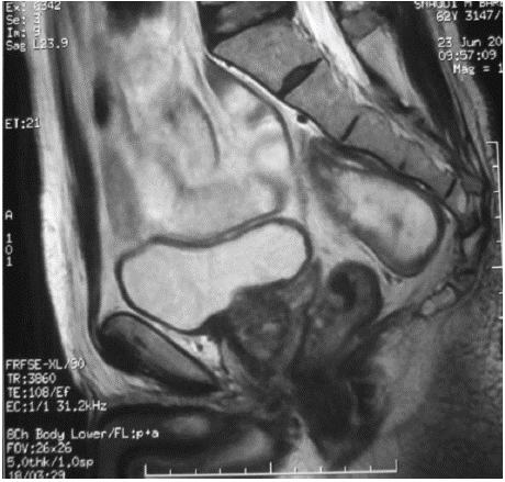
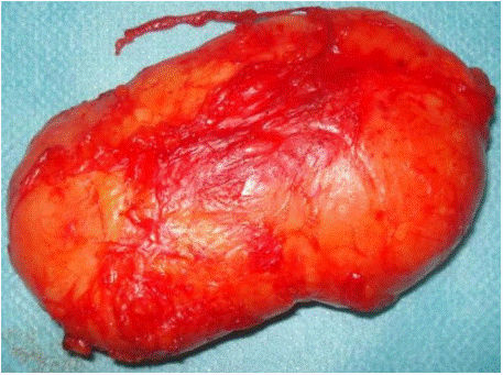
A 69-year-old male patient with a history of type II diabetes 3 years, hypertension, and a stage III LMNH (non-Hodgkin's malignant lymphoma), for which he was receiving CHOP protocol chemotherapy. A control thoracic-abdominal-pelvic CT scan revealed a well-limited, presacral hypodense mass measuring. 70 x 30 mm, with no evidence of extension to neighboring tissues. Pelvic MRI revealed a T1 hypodense mass, isointense on diffusion and T2 sequences, with a necrotic center, well limited, of tissue structure and presacral localization lateralized to the right, oval in shape and measuring 71 x 32 mm (Figure1). The mass extended to the second sacral vertebra, with no invasion of neighboring organs. The surgical approach chosen was an abdominal laparotomy, with on-block tumor resection (Figure2). The anatomopathological analysis confirmed the diagnosis of Ganglioneuroma, with no histological evidence of malignancy. Following surgery, the patient was asymptomatic and showed no signs of recurrence, and was in remission from his non- Hodgkin's lymphoma.

Figure 1: Sagittal section of pelvic MRI showing the presacral lesion.

Figure 2: Peroperative image showing the resected tumor.
Case 2
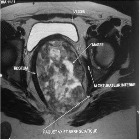
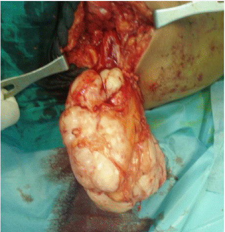
A 25-year-old patient, with no particular pathological history, complained of dyspareunia that persisted for two months, prompting her consultation in the gynecology department. A speculum examination revealed severe pain and compression of the posterior vaginal wall. Pelvic ultrasonography revealed a large, heterogeneous collection, but did not specify its origin. Pelvic MRI confirmed the existence of a left pelvic heterogeneous lesion in T1 hyposignal and T2 hypersignal, with slight progressive enhancement after injection of contrast medium, extending 130 mm in height and measuring 27 x 116 mm in perpendicular diameters (Figure3). The tumor mass was located in the left ischial-anal fossa, compressing the bladder and vagina anteriorly without parietal extension, and pushing the lower rectum, anus, and levator muscles medially. A perineal approach was conducted, with a vertical incision opposite the left ischial-anal fossa. Exploration revealed a voluminous, whitish, encapsulated mass extending high up into the pelvis, with a firm consistency that did not invade any neighboring organs. The mass was resected without capsular invasion (Figure 4). The histological diagnosis was Fibromatosis. Following the operation, and after 4 months of monitoring, the patient was symptomatic and showed no signs of recurrence.

Figure 3: Transversal section of pelvic MRI showing the lesion.

Figure 4: Peroperative image showing the resected mass.
Discussion
The reported cases present two retrorectal tumors, a rare type of tumor often challenging to diagnose due to their nonspecific or asymptomatic nature. Retrorectal tumors are a heterogeneous group of neoplasms that occur in a variety of age groups, but the presentations in these cases reveal distinct contrasts in age, symptoms, imaging characteristics, and surgical management.
Retrorectal neoplasms are uncommon, with an estimated incidence of approximately 1 in 40,000 hospital admissions. These tumors often affect middle-aged adults, though they can occur across a wide range of ages [1]. The notable age difference between the two presented cases, involving a 69-year-old male and a 25-year-old female, underscores the diversity of patients who may develop these tumors [2]. While retrorectal neoplasms have been documented in younger individuals, the majority of cases occur in older adults, rendering our young adult presentation relatively rare [3]. Patient age may also influence tumor characteristics, prognosis, and treatment approaches. Retrorectal tumors are infrequent and, when present, are frequently asymptomatic or exhibit vague symptoms, which can contribute to diagnostic challenges [4]. The first case represents a classic example, where the tumor was incidentally discovered during a follow-up CT scan for lymphoma treatment, despite its considerable size of 71 x 32 mm [5]. In contrast, the second case involved mild symptoms, such as dyspareunia, which prompted the patient to seek medical attention; however, even symptomatic cases like this can present in a nonspecific manner, leading to delayed diagnosis [6]. Studies suggest that up to 50% of retrorectal tumors are asymptomatic, with symptoms typically arising only when the mass compresses adjacent structures [7].
Thorough preoperative planning is essential to determine the most appropriate surgical approach for optimal tumor excision. Computed tomography and magnetic resonance imaging represent the premier preoperative diagnostic modalities [8]. While CT is frequently the initial diagnostic study, it may not consistently differentiate between benign and malignant lesions [9]. In contrast, MRI is the preferred imaging technique due to its superior diagnostic accuracy; it demonstrates greater effectiveness in mass characterization compared to CT, with reported sensitivity and specificity for malignant disease of 81% and 83%, respectively [10,11].
Surgical resection remains the primary treatment modality for retrorectal tumors, with the choice of abdominal approach, perineal, parasacrococcygeal (Kraske procedure) or a combination depending on the tumor size, location, and contact to adjacent organs [12]. Case 1’s abdominal approach via laparotomy facilitated “on block” resection, which is crucial for large, benign tumors to prevent recurrence [13]. In contrast, Case 2’s perineal approach was chosen to access the ischioanal fossa lesion directly, allowing careful excision without capsular invasion, which is critical in cases of fibromatosis where recurrence is possible if resection is incomplete [14].
Ganglioneuromas are typically non-malignant, with low recurrence rates when completely resected, as evidenced by the patient’s remission and lack of recurrence at follow-up [15]. Fibromatosis, although benign, may recur locally, requiring close postoperative monitoring [16]. Both patients have shown no recurrence to date, aligning with literature that emphasizes the importance of complete resection and follow-up to detect early signs of recurrence [17].
Conclusion
Retrorectal tumors are rare, and the two cases highlight the clinical diversity and diagnostic challenges of retrorectal tumors across different age groups. The role of imaging, especially MRI, in establishing a diagnosis and planning for surgical resection, is crucial. Furthermore, complete surgical resection remains the standard of care to minimize recurrence risk in these rare tumors. The successful outcomes in both cases illustrate the importance of individualized treatment approaches based on tumor type, location, and patient factors.
Author Statement
Conflict of Interest
The author has no conflict of interest to declare. The author alone is responsible for the content of this manuscript.
Consent
Written informed consent was obtained from the patients for publication of these case reports and any accompanying.
Ethical Approval
Ethical approval is not required at our institution for an anonymous case report.
References
- Glasgow SC, Birnbaum EH, Lowney JK, Fleshman JW, Mutch MG. Retrorectal tumors: a case report and review of the literature. International Journal of Colorectal Disease. 2005; 20: 261-265.
- Woodfield JC, Phillips N, Spence RAJ. Retrorectal tumours: A comprehensive review of 73 cases. British Journal of Surgery. 2008; 95: 1261-1267.
- Baek SK, Lee YS. Diagnosis and management of retrorectal tumors. Journal of Surgical Oncology. 2014; 109: 108-113.
- Ghosh J, Eglinton T, Frizelle FA, Watson AJ. Presacral tumours in adults. The Surgeon. 2010; 8: 31-38.
- Lev-Cohain N, Chouillard EK. Retrorectal tumors: A comprehensive review of modern diagnostic imaging techniques. European Journal of Radiology. 2012; 81: 2105-2111.
- Hassan I, Wietfeldt ED, You YN. Surgical approach and management of retrorectal tumors: A 20-year experience. Annals of Surgery. 2009; 249: 1053- 1061.
- Lycklama À, Nijeholt AA, Hoekstra HJ. Primary tumors of the retrorectal space: A report of 24 cases and a review of the literature. European Journal of Surgical Oncology. 2006; 32: 533-539.
- Hosseini-Nik H, Hosseinzadeh K, Bhayana R, Jhaveri KS. MR imaging of the retrorectal- presacral tumours: an algorithmic approach. Abdom. Imaging. 2015; 40: 2630–2644.
- Toh J, Morgan M. Management approach and surgical strategies for retrorectal tumours: a systematic review. Colorectal Dis. 2016; 18: 337–350.
- Yang BL, Gu YF, Shao WJ. Retrorectal tumors in adults: magnetic resonance imaging findings. World J Gastroenterol. 2010; 16: 5822–5829.
- Merchea A, Larson DW. The value of peroperative biopsy in the management of solid presacral tumours. Dis. Colon Rectum. 2013; 56: 756–760.
- Jacobs L, Van Der Meer FJ. Management and surgical resection of retrorectal tumors: A review of the current approach. Surgical Oncology. 2012; 104: 99- 105.
- Niwa H, Miura H, Ito H. Surgical resection of retrorectal tumors: An analysis of approaches and outcomes. Journal of Gastrointestinal Surgery. 2016; 20: 1172-1180.
- Hou D, Deng H. Surgical management of retrorectal tumors in a 10-year retrospective study. Journal of Colorectal Disease. 2018; 10: 151-160.
- Damodar K, Sekaran P. Retrorectal ganglioneuroma: Case report and review of literature on surgical management. Clinical Neurology and Neurosurgery. 2015; 133: 83-86.
- MacGregor T, Oates RD. Retrorectal fibromatosis: A rare presentation in young adults. International Journal of Surgical Case Reports. 2014; 5: 80-83.
- Ferhatoglu MF, Uras C. Long-term outcomes following resection of retrorectal tumors: Analysis of recurrence rates. European Journal of Surgical Oncology. 2020; 46: 1191-1198.