
Case Report
Austin J Clin Case Rep. 2024; 11(7): 1345.
Localized Pigmented Villonodular Synovitis of the Knee Joint: A Case Report and Literature Review
Niu Jing, MD¹; Qiao Xu Bai, MD²; Jike Lu, MD, PhD¹*
¹Departments of Orthopaedics, Beijing United Family Hospital, No 2 Jiangtai Road, Chaoyang District, Beijing, 10016, China.
²Departments of Orthopaedics and Pathology, Beijing United Family Hospital, No 2 Jiangtai Road, Chaoyang District, Beijing, 10016, China.
*Corresponding author: Jike Lu, Department of orthopedic Surgery, Beijing united family hospital, Beijing China. Email: jike.lu@ufh.com.cn
Received: November 18, 2024; Accepted: December 10, 2024; Published: December 17, 2024
Abstract
The incidence rate of PVNS, which is synovium tissue as nodules or pedunculated masses, is low however, the localized type is even less. We reported a case of the unusual properties of LPVNS located in the knee.
A 32-year-old man presented to our clinic with pain and mechanic symptoms in his right knee. Magnetic resonance imaging (MRI) showed an intra-articular mass in the infrapatellar area of the knee adjacent to the Hoffa fat pad close to femoral trochlea groove. The mass was hypo intense in the T1 sequence and heterogeneously hypointense in the T2 sequence as well, which was considered as a local type of tenosynovial giant cell tumor (LPVNS). Excision was carefully performed without penetrating the tumor. The gross appearance of the tumor was yellow-reddish and brown in color. Histopathologic examination revealed pigmented villonodular synovitis of the local type.
Localized Pigmented Villonodular Synovitis (LPVNS) is a very rare disease and it is silent and insidious, symptoms are non-specific, nevertheless, differentiation diagnosis should be keeping this entity in mind.
Even though the LPVNS of the knee is an uncommon intra-articular presenttion, overlook this lesion based on imaging findings is an import approach for diagnosis of LPVNS, and arthroscopy assisted excision is an option for management of the LPVNS if doing it appropriately.
Keywords: Pigmented Villonodular Synovitis; Synovial Tumor-like Lesions; Knee pain; Intra-articular Mass
Introduction
Jaffe et al coined the term “PVNS” for the first time in 1941 [1]. Pigmented Villonodular Synovitis (PVNS) most frequently occurs in the knee as a proliferative phenomenon that involves the synovial membrane [2,3]. It is considered a benign lesion of unknown origins. Some authors propose that it is probably caused by inflammation, trauma, toxins, allergies, clonal chromosomal abnormalities, or aneuploidy [4,5].
The disease can be classified into two types: diffuse and localized. The diffuse form (DPVNS), as the name indicates, attacks practically the entire synovial cellular membrane of the affected joint. The localized form (LPVNS) is characterized as a nodule or pedunculated mass. This type is generally a solitary mass of pedunculated or, much less frequently, 2–3 nodules yellowish-brown in color [6-10]. When LPVNS affects the knee, it is generally located in the anterior compartment [6,7].
The incidence rate of PVNS is estimated to be 1.8 per million people—the localized type is just one-quarter of that [11].
Significant swelling with pain at the joint is a characteristic presentation of the patient complaints. Heat and tenderness at and near the joint, limited movement of the joint, locking or catching of the joint popping of the joint, hemarthrosis of blood collects in the joint are common clinical presentations.
Approximately 85% of PVNS occurs in the fingers, whereas 12% of the tumors are located in the knee, elbow, hip, and ankle [12]. PVNS can occur at any age, although it is most common between the ages of 30 and 50, with a female preponderance of 2:1 [13].
Case Report
A 32-year-old man entered our clinic with pain and discomfort in his right knee. The pain had persisted for 3 months, after fell over and twisted his right knee while running. He was recalled that the knee cap was subluxed laterally but reduced spontaneously. He complained the knee was not functioning properly due to catching locking symptoms and restriction of the range of movements. He was difficult to climb stairs and to do squatting activity. Physical examination revealed a painful knee with large effusion of the joint and significantly reduced Range of Movement (ROM), the flexion of the knee was hardly reaching 90 degrees, and Fixed Flexion Deformity (FFD) was 25 degrees. There was pain in flexion and extension, while the patellar tendon was contracted in extension. Lateral and medial McMurray tests, Lachman test was negative. Routine laboratory tests including ESR, CRP, and serum uric acid levels were normal.
Radiographs showed no bony pathology (Figure 1). Magnetic Resonance Imaging (MRI) showed an intra-articular mass of 5× 3 × 1.5 cm in the infrapatellar region of the knee adjacent to the Hoffafat pad. The mass was hypointense in the T1 sequence (Figure 2) and heterogeneously low or intermediate signal in the T2 sequence (Figure 3), which was considered a LPVNS. Based on these findings, arthroscopy-assisted excision of the mass was decided. Under general anesthesia, arthroscopically surgery was performed. Standard portal stabbing incisions were made but medial portal incision was extended longer as required for removal of a large 5 cm x3.5 cm x2cm PVNS mass that is characterized by a single discrete or pedunculated nodule (Figure 5). Removal of all affected parts of the synovium is critical for preventing recurrence. The joint effusion was drained and send for crystal exam in laboratory. Lateral femoral condyle and lateral facet patellar chondral lesion of grade III to IV was seen and abrased with a chondrotone for chondroplasty. A hypoplastic trochlea was identified arthroscopically. The patellar was sitting laterally with subluxation, subsequently lateral retinaculum arthroscopically release was performed with a Radiofrequency (RF). The patellar femoral tracking was checked which was back to normal after lateral release, therefore, no MPFL reconstruction was performed.
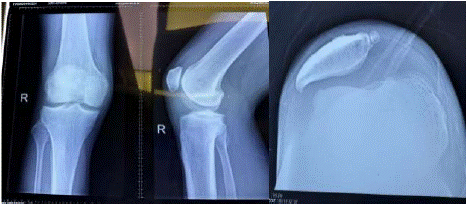
    Figure 1: The lateral radiograph of the knee demonstrates no bony pathology
intra-articularly but he had previous patellar dislocations, patellofemoral joint
instability exists with patellar shifting and tilting laterally with medial facet old
fractured fragment indicating his localized PVNS might be related to his knee
injuries and recurrently subluxation patella.
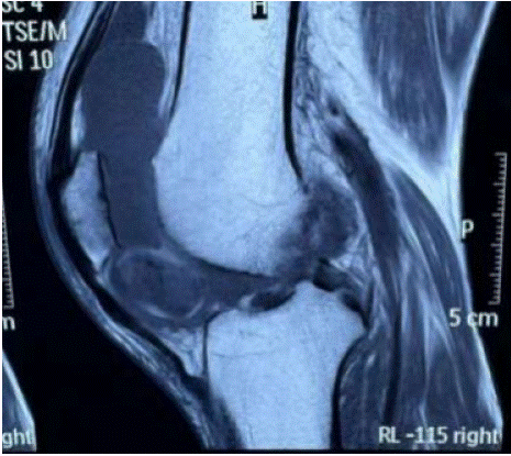
    Figure 2: Sagittal T1-weighted image, arrow shows hypointense mass
located in Hoffa fat pad (A); Axial view showing the mass sitting close to
inferior trochlea groove (B).
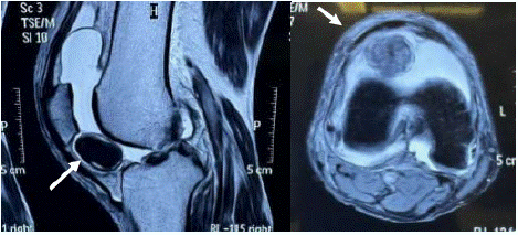
    Figure 3: Sagittal T2-weighted image, the arrow shows heterogeneously
hypointense mass located in Hoffa fat pad.
The mass was identified in close proximity of the Hoffa fat pad. Excision was carefully performed without penetrating the tumor, and the gross appearance of the tumor was yellowish-red and brown in color (Figure 4). A histopathologic examination revealed LPVNS (Figure 6,7). The patient was pain free 3 weeks after surgery. After 12 months follow up, no recurrence had been detected.
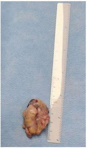
    Figure 4: Gross examination of the tumor mass: The yellowish-red and
patchy brown colored macroscopic appearance of the tumor was noted.
Sizing 5.5 × 3.5 × 2 cm.
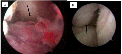
    Figure 5: Arthroscopically excision of the PVNS nodule which was adhearing
on tibia and ACL (arrow), cartilage worn out over lateral femoral condyle and
PF joint (arrow).
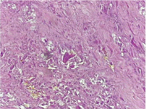
    Figure 6: Histopathologic appearance of the excised lesion showed the
tumor is composed of polymorphous population of epithelioid cells with
histiocytic appearance, macrophages, and osteoclast-like giant cells with
extensive hyalinised necrosis, focal hemosiderin deposits.
Discussion
In this case presentation, we described the clinical and radiologic features of LPVNS and explained the rationale for treatment using arthroscopy-assisted-excision. A comprehensive literature search of localized tenosynovial giant cell tumor or localized pigmented villonodular synovitis of the knee since 1996 was carried out, and 20 articles with 42 cases were found from PubMed search [10,11,14-31]. Only ten articles with ten cases involved the infrapatellar fat pads, excluding two pediatric patients [10,14,15,17-21,23,28]. The ten articles were analyzed to compare the quality of the evidence and the didactic features of the articles. History of trauma is noted in up to 56% of patients with PVNS due to bleeding in joint with subsequent development of hemosiderin deposits and inflammatory markers causing joint destruction. Our case of large localized PVNS which most likely caused by a reactive granulomatous (local) formation rather than a potentially neoplastic formation. From the cases history, most likely repetitive injuries and degenerative joint lesions may be the cause of the reactive granulomatous formation.
It is still debated whether the treatment of LVPNS should be arthrotomy or arthroscopy. There is no consensus on it. Considering the basic principles of tumor surgery, open surgery seems to be more feasible than arthroscopy. This is because it has the advantage of excision of the tumor from the knee without grasping the tumor mass which might be potentially seeding to the joint.
Although the total recurrence rate of PVNS varied from 7% to 44% [32,33], one study reported a rate of 18% after arthroscopic treatment of LPVN within a 6–9 month follow-up [34]. The size of these tumors is the concerns for arthroscopic excision but we had extended portal incision big enough for taking out in one piece through the arthroscopic portal Moreover, meticulously excision of the PVNS nodule in one piece without seeding or piece-meal way to reduce recurrence rate. At the same, debridement of the joint all debris and synovium tissues thoroughly under arthroscopically approach is critical.
In summary, even though LPVNS of the knee is an uncommon intra-articular phenomenon, orthopedic surgeons should not overlook this lesion based on imaging findings, and arthroscopyassisted excision is one of treatment options. However, if the tumor does not appear to come out of the arthroscopic portal in one piece, extension of portal incision should not be hesitated.
References
- Jaffe HL, Lichtenstein L, Sutro CJ. Pigmented villonodular synovitis, bursitis and tenosynovitis. Arch Pathol. 1941; 31: 731–65.
- Byers PD, Cotton RE, Deacon OW, Lowy M, Newman PH, Sissons HA, et al. The diagnosis and treatment of pigmented villonodular synovitis. J Bone Joint Surg Br. 1968; 50: 290–305.
- Granowitz SP, D’Antonio J, Mankin HL. The pathogenesis and long-term end results of pigmenvillonodular synovitis. Clin Orthop. 1976; 114: 335–51.
- Reilly KE, Stern PJ, Dale JA. Recurrent giant cell tumors of the tendon sheath. J Hand Surg. 1999; 24: 1298–302.
- Abdul-Karim FW, El-Naggar AK, Joyce MJ, Makley JT, Carter JR. Diffuse and localized tenosynovial giant cell tumor and pigmented villonodular synovitis:A clinicopathologic and flow cytometric DNA analysis. Hum Pathol. 1992; 23: 729–35.
- Rao AS, Vigorita VJ. Pigmented villonodular synovitis (giant-cell tumor of the tendon sheath and synovial membrane). A review of eighty-one cases. J Bone Joint Surg Am. 1984; 66: 76–94.
- Flandry F, Hughston JC. Pigmented villonodular synovitis. J Bone Joint Surg Am. 1987; 69: 942–949.
- Ogilvie-Harris DJ, McLean J, Zarnett ME. Pigmented villonodular synovitis of the knee. The total arthroscopic synovectomy, partial arthroscopic synovectomy, and arthroscopic local excision. J Bone Joint Surg Am. 1992; 74: 119–23.
- Beguin J, Locker B, Vielpeau C, Souquieres G. Pigmented villonodular synovitis of the knee:Results from 13 cases. Arthroscopy. 1989; 5: 62–4.
- Kim SJ, Shin SJ, Choi NH, Choo ET. Arthroscopic treatment for localized pigmented villonodular synovitis of the knee. Clin Orthop. 2000; 379: 224–30.
- De Carvalho Godoy FA, Faustino CAC, Meneses CS, Nishi ST, Góes CEG, do Canto AL. Localized pigmented villonodular synovitis: Case report. Rev Bras Ortop. 2015; 46: 468–71.
- Fletcher CD, Unni KK, Mertens F. In: World Health Organization Classification of Tumors, Pathology and Genetics of Tumors of Soft Tissue and Bone. Lyon: IARC Press; 2002. Giant cell tumour of tendon sheath. 2002: 110–1.
- Ushijima M, Hashimoto H, Tsuneyoshi M, Enjoji M. Giant cell tumor of the tendon sheath (nodular tenosynovitis): A study of 207 cases to compare the large joint group with the common digit group. Cancer. 1986; 57: 875–84.
- Lucas DR. Tenosynovial giant cell tumor:Case report and review. Arch Pathol Lab Med. 2012; 136: 901–6.
- Beytemür O, Albay C, Tetikkurt US, Oncü M, Baran MA, Caglar S, et al. Localized giant cell tenosynovial tumor seen in the knee joint. Case Rep Orthop. 2014; 2014: 840243.
- Camillieri G, Di Sanzo V, Ferretti M, Calderaro C, Calvisi V. Intra-articular tenosynovialgiant cell tumor arising from the posterior cruciate lig ament. Orthopedics. 2012; 35: e1116–8.
- Ghnaimat M, Alodat M, Aljazazi M, Al-Zaben R, Alshwabkah J. Giant cell tumor of tendon sheath in the knee. Electron Physician. 2016; 25: 2807–9.
- Atik OS, Bozkurt HH, Özcan E, Bahadir B, Uçar M, Ögüt B, et al. Localized pigmented villonodular synovitis in a child knee. Eklem Hastalik Cerrahisi. 2017; 28: 46–9.
- Asik M, Erlap L, Altinel L, Cetik O. Localized pigmented villonodular synovitis of the knee. Arthroscopy. 2001; 17: E23.
- Sun C, Sheng W, Yu H, Han J. Giant cell tumor of the tendon sheath:A rare case in the left knee of a 15-year-old boy. Oncol Lett. 2012; 3: 718–20.
- Flevas DA, Karagiannis AA, Patsea ED, Kontogeorgakos VA, Chouliaras VT. Arthroscopic removal of tenosynovial giant-cell tumors of the cruciate ligaments. presentation of two cases. J Orthop Case Rep. 2021; 11: 23–7.
- Kim RS, Kang JS, Jung JH, Park SW, Park IS, Sun SH. Clustered localized pigmented villonodular synovitis. Arthroscopy. 2005; 21: 761.
- Abdalla RJ, Cohen M, Nóbrega J, Forgas A. Synovial giant cell tumor of the knee. Rev Bras Ortop. 2015; 44: 437–40.
- Tu NV, Quyen NVS, Ngoc MH, Trung HP, Nguyen BS, Trung DT. Tenosynovial giant cell tumor of cruciate ligament: A case report and review. Int J Surg Case Rep. 2022; 91: 106771.
- Xu Z, Mao P, Chen D, Shi D, Dai J, Yao Y, et al. Tenosynovial giant cell tumor arising from the posterior cruciate ligament: A case report and literature review. Int J Clin Exp Pathol. 2015; 8: 6835–40.
- Sheppard DG, Kim EE, Yasko AW, Ayala A. Giant-cell tumor of the tendon sheath arising from the posterior cruciate ligament of the knee:A case report and review of the literature. Clin Imaging. 1998; 22: 428–30.
- Otsuka Y, Mizuta H, Nakamura E, Kudo S, Inoue S, Takagi K. Tenosynovial giant-cell tumor arising from the anterior cruciate ligament of the knee. Arthroscopy. 1996; 12: 496–9.
- GülençB, Kuyucu E, Yalçin S, Çakir A, Bülbül AM. Arthroscopic excision of tendinous giant cell tumors causing locking in the knee joint. Acta Chir Orthop Traumatol Cech. 2018; 85: 109–12.
- Lee JH, Wang SII. A tenosynovial giant cell tumor arising from femoral attachment of the anterior cruciate ligament. Clin Orthop Surg. 2014; 6: 242–4.
- Agarwala S, Agrawal P, Moonot P, Sobti A. A rare case of giant cell tumour arising from anterior cruciate ligament:Its diagnosis and management. J Clin Orthop Trauma. 2015; 6: 140–3.
- Aksoy B, Ertürer E, Toker S, Seçkin F, Sener B. Tenosynovial giant cell tumour of the posterior cruciate ligament and its arthroscopic treatment. Singapore Med J. 2009; 50: e204–5.
- Lowyck H, De Smet L. Recurrence rate of giant cell tumors of the tendon sheath. Eur J Plast Surg. 2006; 28: 385–8.
- Al-Qattan MM. Giant cell tumours of tendon sheath: classification and recurrence rate. J Hand Surg Br. 2001; 26: 72–5.
- Rhee PC, Sassoon AA, Sayeed SA, Stuart MS, Dahm DL. Arthroscopic treatment of localized pigmented villonodular synovitis: Long-term functional results. Am J Orthop (Belle Mead NJ). 2010; 39: 90–4.