
Case Report
Austin J Clin Case Rep. 2015;2(1): 1065.
Axillary Lymph Node Metastases in Differentiated Thyroid Cancer “An Uncommon Presentation with Clinical Implications”
Singhal N1*, Chaturvedi P2, Joshi P3 and Malik A4
1Surgical Oncology Resident, Tata Memorial Hospital, India
2Department of Head and Neck Surgery, Tata Memorial Hospital, India
*Corresponding author: Singhal N, Mch Surgical Oncology Resident, Tata Memorial Hospital, Mumbai, India
Received: January 05, 2015; Accepted: March 23, 2015; Published: March 30, 2015
Abstract
Differentiated thyroid cancers usually follow an indolent course and in general have a good prognosis. Distant metastases at presentation are not common and if present usually involve the lung, bones and brain. Among the various sites reported axillary lymph node is one of the most uncommon sites. Only 13 cases have been reported in literature so far. Here we report two more cases, including the youngest patient reported till date.
Introduction
Differentiated thyroid cancers constitute more than 90 % of the total thyroid cancers and they usually follow an indolent course with excellent prognosis and long term survival. However a small subset of patients present with advanced stage with distant metastases. Incidence of distant metastases at the time of diagnosis is reported to be less than 2% [1]. The common sites of distant metastases are lung and bones with brain, kidney and skin being the other less common sites. Among all the sites of distant metastases reported axillary lymph node is one of the rare sites. Only 13 cases have been reported in literature so far. Here we report two more cases, including the youngest patient reported till date.
Case 1
19 year old female presented in the outpatient clinic with the history of left neck swelling for 4 years and left axillary swelling since 6 months. She had no other significant complaints or family history. On examination there was a left thyroid lobe swelling 4.5 x 3.5 cm with multiple left level II, III, IV and bilateral supraclavicular lymph nodes, largest of which was 5x5 cm along with a left axillary lymph node mass. She was investigated and underwent a USG guided FNAC of thyroid swelling which was diagnostic of papillary thyroid cancer. Ultrasound of breast revealed only left axillary lymph nodal mass of 4.2x3.8 cm, FNAC of which was consistent with metastases from papillary carcinoma of thyroid. As she presented with bulky cervical nodes, a CT scan was done which showed enlarged mediastinal nodes, multiple sub centimeter nodules in both lungs and multiple enlarged lymph nodes in bilateral supraclavicular and left axillary region all of which showed uptake on the PET scan. She underwent total thyroidectomy with bilateral Selective Neck Dissection (SND II-V) with mediastinal node clearance and left axillary lymph node dissection (level I-II) (Figures 1a,1b &1c).
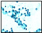
    Figure(a):   Photo of FNAC of thyroid: High power view showing consistent
nuclear features &nuclear groove.
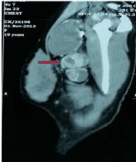
Figure 1(b): CT scan: Showing extensive axillary lymph nodes.
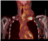
    Figure 1 (c):  PET scan showing uptake in thyroid lobe, mediastinal nodes,
paratracheal nodes and axillary nodes.
Intraoperatively disease was involving the tracheoesophageal groove on left side, encasing the left recurrent laryngeal nerve and going on to esophagus. Decision was taken to leave behind a small amount of disease along the tracheoesophageal groove considering the young age of the patient and morbidity involved with the radical surgery required for R0 resection. The patient recovered well and her final histopathology report was a follicular variant of classical type papillary carcinoma and she underwent radioactive iodine treatment and external beam radiotherapy for the residual disease.
Case 2
45 year old lady underwent right hemithyroidectomy for a right thyroid nodule in 2001 at a local hospital. Histopathology was suggestive of classical variant of papillary thyroid cancer, in view of this she underwent completion thyroidectomy and Left selective neck dissection (II-IV) along with central compartment clearance in 2001 at Tata Memorial hospital. There was no residual tumor identified in the completion thyroidectomy specimen. She was advised a RAI scan post surgery but she defaulted and then presented with the recurrent disease 11 years later. At presentation she had a 10x8x5 cm right supraclavicular mass with palpable right level III and IV lymph nodes and bilateral axillary lymphadenopathy. Serum thyroglobulin was not elevated (0.02) and core biopsy showed no evidence of dedifferentiation. CT scan showed 9x6x5 cm mass from thyroid cartilage to clavicle reaching skin and abutting trachea and common carotid artery. PET-CT revealed two discrete nodules in right lung of 4mm size in upper and lower lobe, along with metabolically active bilateral axillary nodes (Figures 2a, 2b & 2c).

    Figure 2 (a):  Clinical image showing recurrence at neck following first
surgery.
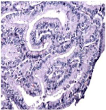
    Figure 2(b):  Microphotograph of resected axillary lymph node mass: tall cell
variant, no area of dedifferentiation.
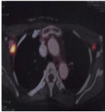
Figure 3(c): PET scan showing uptake in axillary nodes.
She underwent thyroid bed exploration with right neck node clearance with bilateral axillary sampling and DP flap. Histopathology was Tall cell variant with no area of dedifferentiation. Patient was planned for adjuvant Radioiodine treatment but defaulted and had recurrent disease within 7 months. She presented with multiple chest wall nodules largest of which was 6x4 cm along with multiple neck nodes and underwent palliative radiotherapy following which she was again lost to follow up.
Discussion
Axillary lymph node is one of the rarest sites of metastases for thyroid cancers; only 18 cases have been reported in literature so far (Table 1) [2-15]. Of these differentiated thyroid cancers have accounted for 13 cases, with papillary thyroid cancer being the predominant variety accounting for 12 cases and follicular variant contributing a solitary case [13]. Among patients with papillary thyroid ca, 75% (9/12) had poorly differentiated tumor or had an aggressive subtype like tall cell variant. In our series also both the patients were papillary ca with one of them being a tall cell variant.
    Year
    Author
    Age (yrs)
    Sex
    Histology
    Differentiation
    Axillary node appearance
    Metastatic Sites
    Outcome


    2014
    Present study
    19
    F
    Papillary
    Well diff.
    concurrent
    Lung
    Dis Fr 2 mos


    2014
    Present study
    58
    F
    Papillary
    Tall cell.
    Recurrent 11 yrs
    Lung
    NA


    2013
    Cumings [15]
    56
    F
    Papillary
    Well diff.
    Recurrent 8 yrs
    Lung, bone
    ALD


    2013
    Cumings 15
    59
    M
    Medullary
    NA
    Recurrent 3 mo
    None
    Dis Fr 9 mos


    2012
    Machado14
    71
    M
    Papillary
    Well Diff.
    Recurrent 7 yrs
    lung
    ALD 8 months


    2012
    Chiofalo et al. [13]
    65
    M
    Follicular
    Hurthle cell Signet cell
    concurrent
    Multiple bone
    ALD 1year


    2011
    Krishnamurthy et al. [12]
    64
    F
    Papillary
    Tall cell
    Recurrent 6 yrs
    None
    Dis Fr 6 months


    2009
    Kepenecki [11]
    63
    F
    Papillary
    Well diff.
    concurrent
    None
    NA


    2009
    Angeles- Angeles [10]
    58
    F
    Papillary
    Insular
    Recurrent 17 yrs
    Breast
    NA


    2007
    Nakayama [9]
    21
    M
    Papillary
    Partially poorly
    Concurrent
    Lung
    ALD


    2006
    Ers et al. [8]
    62
    F
    Papillary
    NA
    Recurrent 5 yrs
    None
    Dis fr 10 yrs


    2004
    Koike et al. [7]
    51
    M
    Papillary
    Partially poorly
    Recurrent 5 yrs
    Multiple
    Dead


    2003
    Lal [6]
    65
    M
    Papillary
    Poor
    Recurrent 41 yrs
    Multiple
    Dead


    2003
    Lal [6]
    59
    M
    Medullary
    Poor
    concurrent
    Multiple
    ALD


    2003
    Lal [6]
    45
    M
    Papillary
    Poor
    concurrent
    Multiple
    Dead


    2002
    Minagawa et al. [5]
    52
    M
    Mucoepidermoid
    NA
    concurrent
    Lung, vertebra
    Dead


    1998
    Chen et al. [4]
    66
    F
    Papillary
    Well different
    concurrent
    None
    NA


    1996
    Ueda et al. [3]
    45
    F
    Papillary
    NA
    Recurrent 7 yrs
    None
    NA


    1993
    Mizukami et al. [2]
    58
    M
    Adeno Ca (Mucin producing)
    poorly
    Recurrent 7 mos
    None
    Dis Free
Table 1: Table showing the outcomes at different papillary stages.
Mean age of presentation is 55.6 yrs (Range 21-71 yrs). Thus it is mainly seen in late middle or elderly age, however in our series a 19 year old girl presented with axillary metastases which to best of our knowledge is the youngest case ever which has clinical implications since majority of these patients would be subjected to RAI therapy which can cause significant acute and delayed effects including secondary malignancies.
Although thyroid cancers are more common in females, with regards to axillary metastases no sex predilection has been seen with male and female almost equally represented (F- 10 M-8). Among differentiated cancers also this relationship is maintained (F-7 M-6). In our series both the patients were females. About half of these patients presented with concurrent axillary lymph node metastases along with primary. In our series also one patient presented with concurrent axillary metastases while the other one presented in the recurrent disease setting.
All patients reported in literature [2-15] had in common extensive cervical lymph node metastases which can be substantiated by the fact that bulky lymph nodal mass especially in lower cervical region can lead to obstruction of cervical lymphatic vessels and thereby potentiate retrograde lymphatic flow to axillary nodes. Also almost all of them (90%) had at least one of the poor prognostic features (viz. age > 45 yrs, T > 4 cm, presence of multiple sites of distant metastases). This similarities are replicated in our series too as both of our patients had extensive cervical lymph node and lung metastases. Thus, presence of extensive cervical lymph nodes and distant metastases should alert a clinician to look out for axillary nodes as a potential site harboring metastatic disease.
With regards to management of this rare subset of patients there are no formal guidelines as most of the literature concerning advanced differentiated thyroid cancer pertains to laryngotracheal and esophageal invasion. The consensus is to resect and achieve an R0 resection at these sites. Results have been mostly extrapolated from these and till now there are only 7 cases in literature, reported to have undergone axillary dissection [12-15]. Of these 7 patients 2 are disease free, 2 patients are alive with disease while outcomes have not been reported for the remaining 3 patients. In our series one of the patients has just finished treatment for her disease while the other lady had a recurrence within 7 months of surgery; this result should be interpreted with caution taking in to perspective the very advanced nature of her disease along with non adherence to the planned adjuvant treatment.
Conclusion
Thyroid cancer metastasis to axillary lymph nodes is rare; however it has got clinical implications. It should be considered in the differential diagnoses of axillary masses in the presence of thyroid malignancy. Also during follow up, in the setting of persistently raised serum thyroglobulin level and a negative RAI scan clinicians should keep this possibility in mind. Surgical resection with axillary dissection is associated with improved outcomes, but the prognosis is usually ominous.
References
- Nixon IJ, Whitcher MM, Palmer FL, Tuttle RM, Shaha AR, Shah JP, et al. Impact of distant metastases at presentation on prognosis in patients with differentiated carcinoma of the thyroid gland .Thyroid. 2012; 22: 884-889.
- Mizukami Y, Nakajima H, Annen Y, Michigishi T, Nonomura A, Nakamura S. Mucin producing poorly differentiated adenocarcinoma of the thyroid: A case report. PatholRes Pract. 1993; 189: 608-612.
- Ueda S, Takahashi H, Meihe T, Nakata T. A case of axillary lymph nodes recurrence of papillary carcinoma 7 years after the operation. J Jpn Surg Assoc. 1996; 57: 2934-2937.
- Chen SW, Bennett G, Price J. Axillary lymph node calcification due to metastatic papillary carcinoma. Australas Radiol. 1998; 42: 241-243.
- Minagawa A, Iitaka M, Suzuki M, Yasuda S, Kameyama K, Shimada S, et al. A case of primary mucoepidermoid carcinoma of the thyroid: molecular evidence of its origin. Clin Endocrinol. 2002; 57: 551-556.
- Lal G, Ituarte P, Duh QY, Clark O. The axilla as a rare site of metastatic thyroid cancer with ominous implications (abstract only). 74th Annual meeting of ATA. 2003.
- Koike K, Fujii T, Yanaga H, Nakagawa S, Yokoyama G, Yahara T, et al. Axillary lymph node recurrence of papillary thyroid microcarcinoma: report of a case. Surg Today. 2004; 34: 440-443.
- Ers V, Galant C, Malaise J, Rahier J, Daumerie C. Axillary lymph node metastasis in recurrence of papillary thyroid carcinoma. A case report. Wein Klin Wochenschr. 2006; 118: 124-127.
- Nakayama H, Wada N, Masudo Y, Rino Y. Axillary lymph node metastasis from papillary thyroid carcinoma. Report of a case. Surg Today. 2007; 37: 311-315.
- Angeles-Angeles A, Chable-Montero F, Martinez-Benitez B, Albores-Saavedra J. Unusual metastases of papillary thyroid carcinoma: Report of 2 cases. Ann Diag Pathol. 2009; 13: 189-196.
- Kepenecki I, Demirkan A, Cakmak A, Tug T, Ekinci C. Axillary lymph node metastasis as a late manifestation of papillary carcinoma. Thyroid. 2009; 19: 417-419.
- Krishnamurthy A, Vaidhyanathan A. Axillary lymphnode metastasis in papillary carcinoma: report of a case and review of the literature. J Canc Res Ther. 2011; 7: 220-222.
- Chiofalo MG, Losito NS, Fulciniti F, Setola SV, Tommaselli A, Marone U, et al. Axillary node metastasis from differentiated thyroid carcinoma with Hurthle and signet cell differentiation. A case of disseminated thyroid cancer with peculiar histologic findings. BMC Cancer. 2012; 12: 55.
- Machado NO, Chopra PJ, Raouf ZR,Khatib NJ, Sinnakirouchenan. Axillary lymph node metastasis from Thyroid malignancy. Unusual presentation with ominous implications. Br J Med & Med Res. 2013; 3: 1634-1645.
- Cummings AL, Goldfard M. Thyroid carcinoma metastases to axillary lymph nodes: report of two rare cases of papillary and medullary thyroid carcinoma and literature review. Endocr Pract. 2014; 20: e34-e37.