
Case Report
Austin J Clin Case Rep. 2015;2(1): 1067.
Leiomyoma of Oral Cavity in a Young Child
Subramanya Sharma S1*, Ramakrishnan K2, Vijayalakshmi D2 and Saravanan C3
1Department of Oral & Maxillofacial Surgery, Adhiparasakthi Dental College & Hospital, India
2Department of Oral Pathology & Microbiology, Adhiparasakthi Dental College & Hospital, India
3Department of Oral & Maxillofacial Surgery, SRM Dental College & Hospital, India
*Corresponding author: Subramanya Sharma S, Department of Oral & Maxillofacial Surgery, Adhiparasakthi Dental College & Hospital, Melmaruvathur, Tamilnadu, India
Received: December 11, 2014; Accepted: March 23, 2015; Published: April 16, 2015
Abstract
Leiomyoma is a slow-growing, benign tumor of smooth muscle origin presenting clinically as a soft tissue tumor. It occurs more commonly in the gastrointestinal tract, skin, uterus and the subcutaneous tissues. Oral leiomyomas are quite rare. Reported tumors have commonly presented in the lips, tongue, buccal mucosa, palate and mandible. Most of the reported cases are in the 4th and 5th decades of life with very few cases reported in paediatric age group. Most of the lesions are small, asymptomatic and present as a smooth elevated area with pigmentation. We present a case report of an 8-yearold Indian (Asian) boy with a large, exophytic mass in the lingual aspect of the mandible. The size, the site and the age at presentation makes this a rare case worth to be recorded.
Keywords: Smooth muscle; Leiomyoma; Immunohistochemistry
Introduction
Leiomyoma is a benign neoplasm of smooth muscle origin. General absence of smooth muscles in the oral region makes it a rare tumor. Several histological variants described are; Solid Leiomyoma, vascular leiomyoma and Epithelioid variety [1,2]. Vascular leiomyoma, otherwise known as angiomyoma is the most common variant reported in the oral cavity [2,3]. Leiomyoma shows a male predilection of 1.4: 1. It usually presents itself around the 4th to 5th decades of life. The common oral sites are lips, tongue and buccal mucosa. Various other sites including the palate, gingiva and the mandible have been reported [4-6]. Incidence of malignant transformation has not been reported [1,2,7].
We report the case of a male child, who presented with an Oral Leiomyoma.
Case Presentation
An 8-year-old Indian (Asian) boy reported to the out-patient clinic of the Department of Oral & Maxillofacial Surgery with the complaint of a large mass on the lingual aspect of his right mandible in relation to the molar region. The lesion has been present since 6 months and has been growing gradually. It got traumatized frequently during mastication, associated with minor bleeding episodes.
Clinical examination revealed a painless, quite large, solitary, pedunculated, exophytic mass with an irregular, surface in the traumatized areas and other areas were pale pink in color (Figure 1). It measured about 3 x 4 cm in size extending anteriorly from the deciduous first molar to the retromolar area posteriorly on the right lingual aspect of alveolar mucosa and gingiva. Superiorly, the lesion extended to the line of occlusion and inferiorly upto the floor of mouth. Medially the lesion was adherent to the lingual aspect of mandible. The lesion was firm in consistency. No regional lymphadenopathy was evident. Differential diagnoses for the peripheral, oral exophytic lesion are Inflammatory Hyperplasias and peripheral benign mesenchymal tumors. An excisional biopsy was done and the specimen was sent to the Oral Pathology Department (Figure 2).
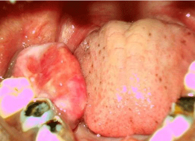
Figure 1: Intraoral lesion.
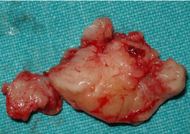
Figure 2: Excised specimen.
The routine H & E section of the excised lesion showed a parakeratinized stratified squamous surface epithelium associated with the underlying tumor parenchyma which was composed of interlacing fascicles of spindle cells with eosinophilic cytoplasm and elongated, pale staining, vesicular nuclei (Figure 3). The nuclei were blunt ended, typical of smooth muscle nuclei (Figure 4). The presence of few compressed slit-like vessels was evident. No mitotic activity was seen. The lesion was classified as a benign spindle cell neoplasm and histological differential diagnosis include Leiomyoma, Myofibroblastic tumor, Fibrous histiocytoma and Neurilemoma. The absence of histiocytes, multinucleated giant cells and absence of Antoni type A, type B, verocay bodies differentiated the lesion from Fibrous Histiocytoma and Neurilemoma respectively.
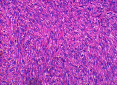
    Figure 3:  Photomicrograph showing interlacing fascicles of spindle cells with
blunt ended nuclei.
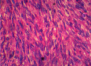
Figure 4: Photomicrograph showing spindle cells with blunt ended nuclei.
Immunohistochemistry (IHC) studies were performed for further differentiating the spindle cell neoplasms with the following markers namely S-100, SMA (Smooth Muscle Actin), Muscle specific Actin (HHF-35), Desmin, Caldesmon, Vimentin, Calponin, and Ki67. Strong positive expression was noted for SMA (Figure 5), Desmin (Figure 6) and Caldesmon (Figure 7). S-100, Calponin and HHF-35 were negatively expressed. Ki -67 labelling index was insignificant. Thus the tumor was diagnosed as Leiomyoma.
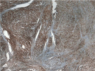
Figure 5: Photomicrograph showing diffuse positivity for SMA.
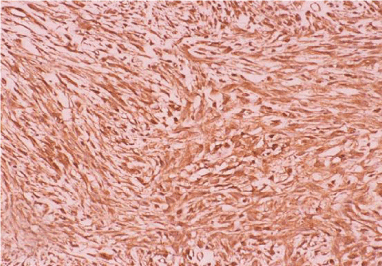
Figure 6: Photomicrograph showing positivity for Desmin.
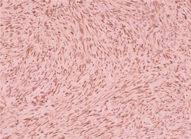
Figure 7: Photomicrograph showing positivity for Caldesmon.
Discussion
Oral leiomyoma accounts for less than 1% of all leiomyomas. Oral leiomyoma is hypothesized to originate from vascular smooth muscles and excretory ducts of salivary glands. In the oral cavity, vascular or angiomyoma is the most common variant followed by the solid variant. Epithelioid variant (Leiomyoblastoma) is said to be extremely rare accounting for less than 1% [8]. In the oral cavity it rarely attains a huge size with our case possibly the largest reported [2,3,9].
Histopathologically leiomyoma may be confused with other spindle cell neoplasms. We suggest that an oral leiomyoma, in special occasions such as involving young individuals needs Immunohistochemical stains with a panel of antibodies like Caldesmon, Desmin, HHF-35, Calponin, and Ki-67 to confirm the histopathological diagnosis. The typical cigar-shaped nuclei arranged in whirls and interlacing bundles and the brighter eosinophilic cytoplasm in leiomyoma differentiates it from a Myofibroblastic tumor [1,2,7,10]. Neurilemomas are S-100 positive [11]. Both leiomyomas and myofibromas express SMA [12,13]. Leiomyomas express Desmin and Caldesmon whereas Myofibroblastic tumors are negative [14,15]. Vimentin, HHF-35, Calponin and Ki67 are positive for Myofibroblastic tumors and negative for leiomyomas [16,17]. Therefore, Immunohistochemistry plays a vital role in distinguishing the spindle cell neoplasms.
References
- Rajendran R, Sivapathasundaram B. Shafer’s textbook of Oral Pathology. 5th edn. Elsevier Publishers. 2006.
- Gaitan Cepeda LA, Quezada Rivera D, Rocha FT, Leyva Huerta ER, Mendez Sànchez ER. Vascular leiomyoma of the oral cavity. Clinical, histopathological and immunohistochemical characteristics. Presentation of five cases and review of the literature. Med Oral Patol Oral Cir Bucal. 2008; 13: E483-488.
- Brooks JK, Nikitakis NG, Goodman NJ, Levy BA. Clinicopathology characterization of oral angioleiomyomas. Oral Surg Oral Med Oral Pathol Oral Radiol Endod. 2002; 94: 221-227.
- Luaces Rey R, Lorenzo Franco F, Gomez Oliveira G, Patino Seijas B, Guitiàn D, LòpezCedrÙn Cembranos JL. Oral leiomyoma in retromolar trigone. A case report. Med Oral Patol Oral Cir Bucal. 2007; 12: E53-55.
- Bhattacharyya I, Don-John S, Cohen DM, Ellis GL, Bavitz JB, Gillham LL. Granular cell leiomyoma of the oral cavity. J Oral Maxillofac Pathol. 2006; 102: 353-359.
- Koca H, Guneri P, Cetingul E, Onal T. A very rare form of leiomyoma: Mandibular angioleiomyoma. Int J Pediatric Otorhinolaryngol. 2006; 1: 110-114.
- Baden E, Doyle JL, Lederman DA. Leiomyoma of the oral cavity: a light microscopic and immunohistochemical study with review of the literature from 1984 to 1992. Eur J Cancer B Oral Oncol. 1994; 30B: 1-7.
- Ioannis KG, Manivel J, Carlos. Epitheliod leiomyomaof the mandible. J Oral Maxillofac Pathol. 1996; 82: 670-673.
- Svane TJ, Smith BR, Consentino BJ, Cundiff EJ, Ceravolo JJ. Oral leiomyomas: Review of the literature and report of a case of palatal angioleiomyomas. J Periodontol. 1986; 57: 433-435.
- Reddy B, Rani BS, Anuradha C, Chandrasekhar P, Shamala R, Lingamaneni K. Leiomyoma of the mandible in a child. J Oral Maxillofac Pathol. 2011; 15: 101-104.
- Weiss SW, Langloss JM, Enzinger FM. Value of S-100 protein in the diagnosis of soft tissue tumors with particular reference to benign and malignant Schwann cell tumors. Lab Invest. 1983; 49: 299-308.
- Aatish Thennavan, Venkadasalapathi Narayanaswamy, Thanvir Mohammed Niazi, Lakshmi Rao, Raghu Radhakrishnan. Infantile Myofibroma Eroding into the Frontal Bone: A Case Report and Review of Its Histopathologic Differential Diagnosis. Hindawi Publishing Corporation. Case Reports in Pediatrics. 2012; 2012: 630804.
- Kaur G, Gondal R. Oral leiomyoma. J Oral Maxillofac Pathol. 2011; 15: 361-362.
- Efraín Alvarez, Laberry MP, Ardila CM. Case Report. Multiple Oral Leiomyomas in an Infant: A Rare Case. Hindawi Publishing Corporation. Case Reports in Dentistry. 2012; 2012: 804305.
- Takahiro Yamanishi, Kaname Sakamoto, Hiroyuki Watanabe, Takaaki Yonaga, Naoki Oishi, Ryohei Katoh, et al. Primary Cervical Leiomyoma with Remarkable Calcification and Ossification. Hindawi Publishing Corporation. Case Reports in Otolaryngology. 2014; 2014: 896275.
- Cargini P, Fidanza F, Facente MV, Sgolastra F, Gatto R, Cutilli T. Gingival Myofibroma. A case report. Eur J Paediatr Dent. 2012; 13: 81-83.
- Brasileiro BF, Martins-Filho PR, Piva MR, da Silva LC, Nonaka CF, Miguel MC. Myofibroma of the oral cavity. A rare spindle cell neoplasm. Med Oral Patol Oral Cir Bucal. 2010; 1:15.