Case Report
Austin J Clin Case Rep. 2015; 2(3): 1075.
Endogenous Klebsiella Pneumoniae Endophthalmitis in a Diabetic Patient
Bazoukis G¹*, Boukas K¹, Fytrakis N¹, Florou K¹, Spiliopoulou A¹, Kaperda A¹, Thrappas J¹, Bazoukis X¹, Bakouli A², Savvanis S¹, Fragkou A¹, Yalouris A¹
¹Department of Internal Medicine, General Hospital of Athens “Elpis”, Greece
²Department of Ophthalmology, General Hospital of Athens “Elpis”, Greece
*Corresponding author: George Bazoukis, Department of Internal Medicine, General Hospital of Athens (Elpis) Greece, Dimitsanas 7, Ambelokipi, Athens, Greece
Received: June 24, 2015; Accepted: September 01, 2015; Published: September 09, 2015
Abstract
Endogenous Klebsiella Pneumoniae endophthalmitis is considered a rare complication of gram negative sepsis. Despite immediate management, the visual outcome of patients with endogenous K. Pneumoniae endophthalmitis is poor ranging from hand motion visualization to evisceration or enucleation of the eye. To the best of our knowledge, we present the first reported case in Greece and among the few reported cases in Europe of a diabetic patient with endogenous K. Pneumoniae endophthalmitis.
Keywords: Endogenous endophthalmitis; Klebsiella Pneumoniae; Leftsided endopthalmitis; Multifocal pneumonia
Abbreviations
EKE: Endogenous Klebsiella Pneumoniae Endopthalmitis
Introduction
Endogenous bacterial endophthalmitis is a rare entity that accounts for 2–15% of all cases of endophthalmitis [1]. Endogenous Klebsiella Pneumoniae endophthalmitis (EKE) is considered a rare complication of gram negative sepsis [2]. A significant increase of EKE in Asia has been reported [2,3]. Although that condition is more prevalent in Asia, cases of K. Pneumoniae liver abscess with endophthalmitis have been also reported in the USA, Australia, Spain, UK and the Middle East [4]. Conditions like intravenous drug abuse, treatment with immunosuppressive agents and diabetes mellitus predispose patients to the disease [5]. The primary sources of EKE infection are suppurative liver disease (68%) or urinary tract infection (16%) [2]. Ten percent of pyogenic K. Pneumoniae liver abscess cases are complicated by EKE [5]. However, the presence of EKE with only bilateral pulmonary infiltrations has rarely been reported [1]. Despite immediate management, the visual outcome in patients with EKE is poor and it ranges from hand motion visualization to evisceration or enucleation of the eye [5].
To the best of our knowledge, we present the first reported case in Greece and among the few reported cases in Europe of a diabetic patient with endogenous K. Pneumoniae endophthalmitis.
Case Presentation
A 55-year-old man from Greece was admitted at the emergency department of our hospital for a 10-day history of fever and cough. He has been empirically treated with Amoxicillin-Clavulanic acid for a week without significant improvement of the symptoms. In addition to that the patient reported redness with concomitant pain and a sudden decrease of the visual acuity of the left eye starting the last 24 hours. He did not mention a recent travel abroad. His medical history included a 6 years history of diabetes mellitus without receiving any medication and hypertension treated with atenolol and irbesartan/hydrochlorothiazide combination.
On admission the patient was febrile. On clinical examination, there was a mild periorbital edema with chemosis of the left eye. The ophthalmological examination showed a significant impairment of the visual acuity limited to “light perception” while the slit lamp examination revealed hypopyon 3,0 mm. The right eye was free of any pathology. Chest x-ray showed the presence of bilateral diffuse lung infiltrates.
Initial blood results showed raised inflammatory markers with white blood cells count (WBC) of 23.900/μL (neutrophil count of 20.600/μL) and C-reactive protein of 18, 20 mg/dl. The random blood glucose levels were 489 mg/dl while the HbA1C was 12, 50%. Other findings were: serum creatinine: 1,3mg/dl, serum sodium: 128mmol/l, serum potassium: 5,0mmol/l, alanine aminotransferase (ALT): 46 U/l, aspartate aminotransferase (AST): 40 U/l and total bilirubin: 1,10mg/dl. The arterial blood gases were within normal values. Our patient was HIV negative and there was no evidence of urinary tract infection.
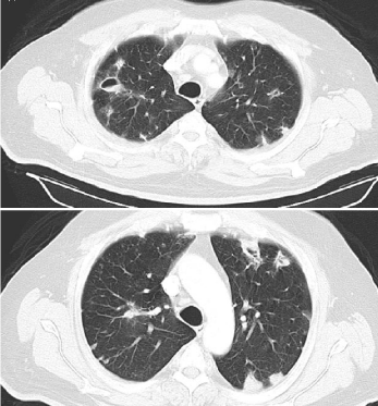
The blood cultures were positive for K. Pneumoniae (Table 1) while the chest computed tomography scan (CT scan) showed multiple cavitary lesions and nodules of various sizes (Figure 1). The abdominal CT scan did not reveal pathological findings. Transthoracic echocardiogram did not show valvular vegetations. As a result the patient was diagnosed with endogenous K. Pneumoniae endophthalmitis secondary to a systemic infection.
    K. Pneumoniae antibiogram


    Antibiotic
    MIC


    Amikacin
    =16


    Ampicillin/Sulbactam
    =8/4


    Ampicillin
    >16


    Cefepime
    =8


    Cefotaxime
    =1


    Cefoxitin
    =8


    Ceftazidime
    =1


    Cefuroxime
    =4


    Ciprofloxacin
    =1


    Colistin
    =2


    Ertapenem
    =0.5


    Gentamicin
    =4


    Imipenem
    =1


    Levofloxacin
    =2


    Meropenem
    =1


    Moxifloxacin
    =0.5


    Nitrofurantoin
    >64


    Piperacillin/Tazobactam
    =16


    Piperacillin
    =16


    Tetracycline
    =4


    Tigecycline
    =1


    Tobramycin
    =4


    Trimethoprim/Sulfamethoxazole
    =2/38
Table 1: Klebsiella Pneumoniae antibiogram.

    Figure 1:  The chest computed tomography revealed multiple cavitary lesions
and nodules of various sizes.
The patient was immediately given intravenous meropenem 2 gr tid and gentamicin 80 mg tid which were initiated for 25 days in total. The blood glucose levels were tightly controlled with short and long acting insulin analogs. After ophthalmologists’ consultation, the patient was given intravitreal injections of amikacin while fortified tobramycin 15mg/ml and ciprofloxacin 0, 3% eye drops were given every hour. The visual acuity was declined to “no light perception” within the next five days despite the improvement of both the pain and hypopyon. Acetazolamide 250 mg qid p.os was initiated for preventing increase in intraocular pressure.
On discharge (day 28), his blood tests revealed WBC: 9.800/μl and C-reactive protein: 2,05mg/dl without impairment of the kidney and liver functions. The visual acuity was preserved in the right eye while the patient had no vision on the left eye.
Discussion
K. pneumoniae, a common human pathogen, is most often associated with pneumonia and urinary tract infections. Liver abscesses with metastatic infections caused by serotypes K1 and K2 of K. Pneumoniae have been reported with increasing frequency in Asia [4]. Extrahepatic infections include bacteriuria, meningitis, endophthalmitis, necrotizing fasciitis, endocarditis, spinal epidural abscesses, septic pulmonary embolism or abscess, septic arthritis, prostate and renal abscesses [2,4,6].
Diabetes mellitus has been identified as the major risk factors to develop EKE [7]. Impaired phagocytosis of capsular serotypes K1 or K2 K. Pneumoniae in type 2 diabetes mellitus patients with poor glycemic control like the patient of our case report may play an important role for the pathogenesis of serious metastatic complications in diabetic patients [8]. Furthermore, the diabetic ocular changes may lead to an increase in the blood-retinal barrier permeability contributing with that way in the higher incidence of EKE in diabetes [5].
Generally, the right eye is affected more often than the left eye, which is probably due to direct blood flow from the heart [9]. Painful ocular swelling, redness, and sudden-onset blurred vision are the classic ocular complaints [7]. In a recent study, investigators showed that both whole live K. Pneumoniae and K. Pneumoniae lipopolysaccharide exert a strong pro-inflammatory effect on retinal epithelial cells, consistent with the clinical presentation of disease [10]. Early diagnosis and intensive intravenous antibiotics are the most critical steps in the treatment of endogenous bacterial endophthalmitis. Vitreous biopsy has a weak diagnostic relevance as it is often negative [11]. Because of blood-retinal barrier, intravitreal injection of antibiotics may be adopted for achieving effective therapeutic drug levels within the vitreous [8]. Ceftriaxone or meropenem with good penetration into vitreous are good choices for intravenous infusion [8,12]. Although in some case series and in accordance to preclinical models, the early intravitreal corticosteroids injection lead to better visual outcomes [2], its use in cases of acute endophthalmitis remains controversial [13].
Despite adequate treatment, EKE has a poor visual outcome due to its extreme virulence and delay in the treatment ranging from hand motion visualization to evisceration or enucleation of the eye [5]. Prognostic factors for adverse visual outcome include the rapid onset of ocular symptoms, unilateral involvement, the presence of hypopyon and panophthalmic involvement (as opposed to posterior focal or diffuse involvement) [14]. Also, unilateral involvement showed to be related with an increased likelihood of having an evisceration [14].
Conclusion
In conclusion, although EKE is more prevalent in Asia, clinicians should be aware of the disease because early recognition and appropriate treatment under ophthalmologists’ consultation are the cornerstones for achieving a better visual outcome.
References
- Yin W, Zhou H1, Li C. Endogenous Klebsiella pneumoniae endophthalmitis. Am J Emerg Med. 2014; 32: 1300.
- Yang CS, Tsai HY, Sung CS, Lin KH, Lee FL, Hsu WM. Endogenous Klebsiella endophthalmitis associated with pyogenic liver abscess. Ophthalmology. 2007; 114: 876-880.
- Kashani AH, Eliott D. The emergence of Klebsiella pneumoniae endogenous endophthalmitis in the USA: basic and clinical advances. J Ophthalmic Inflamm Infect. 2013; 3: 28.
- Abdul-Hamid A, Bailey SJ. Klebsiella pneumoniae liver abscess and endophthalmitis. BMJ Case Rep. 2013; 2013.
- Coburn PS, Wiskur BJ, Christy E, Callegan MC. The diabetic ocular environment facilitates the development of endogenous bacterial endophthalmitis. Invest Ophthalmol Vis Sci. 2012; 53: 7426-7431.
- Chen KJ, Hwang YS, Wang NK, Chao AN. Endogenous Klebsiella pneumoniae endophthalmitis with renal abscess: Report of two cases. Int J Infect Dis. 2010; 14: e429-432.
- Al-Mahmood AM, Al-Binali GY, Alkatan H, Abboud EB, Abu El-Asrar AM. Endogenous endophthalmitis associated with liver abscess caused by Klebsiella pneumoniae. Int Ophthalmol. 2011; 31: 145-148.
- Lin JC, Siu LK, Fung CP, Tsou HH, Wang JJ, Chen CT, et al. Impaired phagocytosis of capsular serotypes K1 or K2 Klebsiella pneumoniae in type 2 diabetes mellitus patients with poor glycemic control. J Clin Endocrinol Metab. 2006; 91: 3084-3087.
- Arcieri ES, Jorge EF, de Abrea Ferreira L. Bilateral endogenous endophthalmitis associated with infective endocarditis: case report. Braz J Infect Dis. 2001; 5: 356-359.
- Pollreisz A, Rafferty B, Kozarov E, Lalla E. Klebsiella pneumoniae induces an inflammatory response in human retinal-pigmented epithelial cells. Biochem Biophys Res Commun. 2012; 418: 33-37.
- Guber J, Saeed MU. Presentation and outcome of a cluster of patients with endogenous endophthalmitis: a case series. Klin Monbl Augenheilkd. 2015; 232: 595-598.
- Schauersberger J, Amon M, Wedrich A, Nepp J, El Menyawi I, Derbolav A, et al. Penetration and decay of meropenem into the human aqueous humor and vitreous. J Ocul Pharmacol Ther. 1999; 15: 439-445.
- Bui DK, Carvounis PE. Evidence for and against intravitreous corticosteroids in addition to intravitreous antibiotics for acute endophthalmitis. Int Ophthalmol Clin. 2014; 54: 215-224.
- Ang M, Jap A, Chee SP. Prognostic factors and outcomes in endogenous Klebsiella pneumoniae endophthalmitis. Am J Ophthalmol. 2011; 151: 338-344.