Case Report
Austin J Clin Case Rep. 2015; 2(3): 1077.
Intramuscular Ganglion Cyst: A Common Lesion in an Unusual Location
Najjar S* and Nasser H
Department of Pathology, Prince Sultan Military Medical City, Saudi Arabia
*Corresponding author: Najjar S, Department of Pathology and Laboratory Medicine, King Faisal Specialist Hospital and Research Center, Prince Sultan Military Medical City, MBC10, Saudi Arabia
Received: July 06, 2015; Accepted: September 05, 2015; Published: September 10, 2015
Abstract
Ganglion cysts represent the most common benign soft tissue lesion of the hands and wrists. When arising in their characteristic locations, their diagnosis and treatment is easy; however, when arising in atypical locations they pose a diagnostic dilemma. We present a case of intramuscular ganglion cyst arising in the extensor digitorum muscle of the left arm. Due to its unusual location and radiological characteristics it was suspected as being an intramuscular myxoma, and was treated with wide excision. We believe that recognizing this entity in the differential diagnosis of intramuscular cystic lesions is important for proper management.
Keywords: Ganglion cyst; Intramuscular cyst; Radiological
Introduction
Ganglion cysts (GCs) are non-neoplastic pseudocysts that have no true epithelial lining and are filled with gelatinous material composed mainly of hyaluronic acid. Their definite etiology is still unknown. They may be secondary to degenerative changes and chronic damage that leads to liquefaction and cyst formation. This is followed by proliferation and formation of a fibrotic and compact wall arising from the surrounding connective tissue [1] The gelatinous material has also been proposed to be produced by injured mesenchymal cells [2].
GCs most commonly originate from joints, particularly the scapholunate joint of the wrist, and to a lesser extent form tendon sheathes in young patients. However, they have been reported in many different sites and so have a variety of clinical presentations depending on their anatomical location.
GCs arising in atypical locations present a diagnostic challenge and might get misdiagnosed. They have been reported to originate from cartilage, nerves, and muscles. Intra articular ganglion cysts of the knee mainly involve the tendon sheath or joint capsule and infrequently the menisci, and anterior and posterior cruciate ligaments [3-5]. Somewhere reported to occur intraosseously in the distal epiphysis of the tibia and from peripheral nerve sheath particularly the common perineal nerve [6]. Involvement of other nerves including the radial, ulnar, median and sciatic nerves have also been reported [7]. Cases of multiple ganglion cysts involving unusual sites like the temporo mandibular joint affecting young patients, the so called cystic ganglion sis, might indicate a genetic susceptibility and give new insights to the pathogenesis of this lesion [8].
Case Presentation
Clinical summary
A 46-year-old male patient, medically free, presented complaining of a left upper arm swelling of six months duration. On physical examination, a bulging well-circumscribed mass was noted in his elbow region. There was no range of motion restriction, weaknesses or paresthesia.
Radiological findings
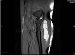
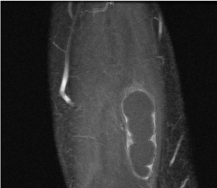
Multiplanar MRI of the left elbow joint showed a multiloculated multicystic lesion at the extensor digitorum muscle from the level of the radial head showing thin peripheral wall and septal enhancement of the mass without internal solid enhancement confirming its cystic nature (Figure 1). The tendons and ligaments around the elbow joint were normal and unaffected. No connection to the elbow joint was identified and the diagnosis of intramuscular myxoma was suggested.

    Figure 1a:  Left elbow joint MRI: hyper intense lobulated mass just below the
elbow joint, coronal T2.

    Figure 1b:  Left elbow joint MRI: Thin peripheral wall and septal enhancement
of the mass, coronal Post gadolinium sequence.
Pathological findings
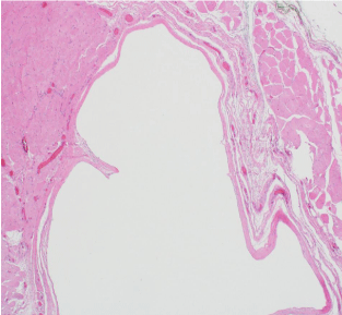
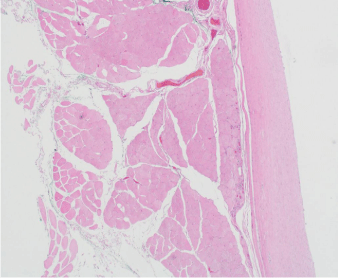
The patient underwent local resection with wide margins and tumor bed excision. The surgical sample demonstrated an oval, well-circumscribed 5.0x2.6x1.5 cm mass with surrounding attached skeletal muscle. Serial sectioning revealed a multiloculated cyst filled with thick viscous gelatinous material. The inner lining was smooth and glistening. Microscopic examination revealed a thin fibrous cyst wall without a true lining, and surrounded by compressed skeletal muscles (Figure 2).

    Figure 2A:  partially septated cyst wall with clear content, surrounded by
skeletal muscle (H&E; 4x magnification).

Figure 2B: Thin fibrous wall of the cyst (H&E; 40x magnification).
Discussion
GCs involving skeletal muscle commonly represent extension from nearby joints they frequently arise in the rotator cuff muscles of the shoulder joint and are strongly associated with rotator cuff tendon tears particularly the supraspinatus. It’s proposed that a tear in the muscle sheath lead to dissection of fluids into the muscle substance leading to the formation of these ganglia [9].
In about 30% of cases ganglion cysts may arise as isolated intramuscular cysts [10]. Muscles reported to be involved include quadriceps, biceps brachial is and gastronomies [11-14]. No cases have been reported in the extensor digitorum muscle to date.
Isolated intra muscular GCs are usually less than 5 cm in maximum diameter, however, a large GC with an11 cm diameter, and interfering with patient’s ambulation is reported in the vastuslateralis muscle [15]. They tend to grow slowly within several months to years. The majority of isolated intramuscular GCs are deep seated but cases presenting as subcutaneous masses has been described [16]. James et al. think that isolated intramuscular GCs are more prevalent than expected due to the fact that the majority of them are asymptomatic [17]. Rare symptomatic cases commonly presented with pain due to compression of nearby nerves or rupture [10,18]. A case of an isolated peroneus muscle GC presented as perineal compartment syndrome [19].
Patients presenting with intramuscular GCs are initially approached with ultrasound examination as it is readily available and noninvasive. Intramuscular GCs appear as hypoechoic or anechoic cysts. Further MRI studies will provide better characterization of the mass, its location and relationship to the surrounding structures. Intramuscular GCs are lobulated multi or unilocular cysts with will defined septa exhibiting homogenous low-signal on T1 weighted images and high-signal on T2 weighted images with Contrast enhancement at its borders [20]. That being said, these radiological features are non-specific and can be seen with other soft tissue neoplasms, particularly those rich with myxoid matrix, like intramuscular myxoma, myxoid neurofibromas and myxoid liposarcomas. Intramuscular myxoma is a benign neoplasm showing sparse bland looking spindle cells in an abundant myxoid background surrounded by a pseudo capsule and is well known to undergo cystic degeneration. It is hypoechoic on ultrasound and have similar low T1 high T2 signal, however, it tends to show more heterogeneous enhancement on MRI [17,21]. Intramuscular myxomas tend to recur after excision due to their deep location, especially if incompletely excised. Myxoidneurofibromais a rare sub type of the classical neurofibroma showing similar cells with tapered nuclei and scanty cytoplasm in a myxoid background. Myxoid liposarcoma, a malignant tumor that requires a wider excision with clear margins, usually presents as a well-circumscribed and hypo cellular tumor showing abundant myxoid background with characteristic delicate plexiform vascular channels and a variably cellular background with round to oval nuclei. It is important to note that a number of soft tissue neoplasms may undergo myxoid degeneration, whether focally or diffusely, and therefore can have similar radiologic features.
The proper diagnosis of such cases is important for appropriate treatment. Simple cysts are treated best with simple excision with a negligible rate of recurrence while synovial cysts are best aspirated.
We report the first GCto arise in the extensor digitorum muscle. It was excised with wide margins, but no complications for the patient. We recommend including GCs in the differential diagnosis list of any intramuscular cystic lesion, especially when demonstrating appropriate radiological features.
References
- Soren A. Pathogenesis and treatment of ganglion. Clin Orthop Relat Res. 1966; 48: 173-179.
- Thornburg LE. Ganglions of the hand and wrist. J Am Acad Orthop Surg. 1999; 7: 231-238.
- Ogawa H, Itokazu M, Ito Y, Fukuta M, Simizu K. An unusual meniscal ganglion cyst that triggered recurrent hemarthrosis of the knee. Arthroscopy. 2006; 22: 455.
- Beaman FD, Peterson JJ. MR imaging of cysts, ganglia, and bursae about the knee. Radiol Clin North Am. 2007; 45: 969-982.
- Fukuda A, Kato K, Sudo A, Uchida A. Ganglion cyst arising from the posterolateral capsule of the knee. J Orthop Sci. 2010; 15: 261-264.
- Sakamoto A, Oda Y, Iwamoto Y. Intraosseous Ganglia: a series of 17 treated cases. Biomed Res Int. 2013; 2013: 462730.
- Kwon OS, Bahk WJ, In Y. Common Peroneal Nerve Palsy Caused by a Ganglion - Case Report. J Korean Orthop Assoc. 2003; 38: 531-534.
- Shinawi M, Hicks J, Guillerman RP, Jones J, Brandt M, Perez M, et al. Multiple ganglion cysts ('cystic ganglionosis'): an unusual presentation in a child. Scand J Rheumatol. 2007; 36: 145-148.
- Kassarjian A, Torriani M, Ouellette H, Palmer WE. Intramuscular rotator cuff cysts: association with tendon tears on MRI and arthroscopy. AJR Am J Roentgenol. 2005; 185: 160-165.
- Kim YJ, Chae SU, Choi BS, Kim JY, Jo HJ. Intramuscular ganglion of the quadriceps femoris. Knee Surg Relat Res. 2013; 25: 40-42.
- Yang SW, Teng HP, Tarng YW, Wong CY. Intramuscular Ganglion Cyst of the Quadriceps Muscle: Report of a Case. Mid Taiwan J Med. 2002; 7: 193-719.
- Rohrich RJ, Rich BK. Intramuscular ganglion of the biceps muscle. Ann Plast Surg. 1994; 33: 432-433.
- Beggs I, Saifuddin A, Limb D. Non-communicating intramuscular ganglia. Eur Radiol. 1998; 8: 1657-1661.
- Jackson L, Namey TC. Ganglion cyst within the quadriceps muscle: evaluation with computed tomography and ultrasound. A case report. Orthopedics. 1987; 10: 1179-1180.
- Vayvada H, Tayfur V, Menderes A, Yilmaz M, Barutcu A. Giant ganglion cyst of the quadriceps femoris tendon. Knee Surg Sports Traumatol Arthrosc. 2003; 11: 260-262.
- Park JJ, Park JG, Lee JB, Kim SJ, Lee SC, Won YH, et al. Intramuscular Ganglion Cyst of the Gastrocnemius Muscle. Korean J Dermatol. 2010; 48: 56-59.
- James SL, Connell DA, Bell J, Saifuddin A. Ganglion cysts at the gastrocnemius origin: a series of ten cases. Skeletal Radiol. 2007; 36: 139-143.
- Park S, Jin W, Chun YS, Park SY, Kim HC, Kim GY, et al. Ruptured intramuscular ganglion cyst in the gastrocnemius medialis muscle: sonographic appearance. J Clin Ultrasound. 2009; 37: 478-481.
- Kang SY, Lee HJ, Lee AH. Intramuscular ganglion of the peroneus muscle mimicking peroneal compartment syndrome. J Korean Orthop Assoc. 2004; 39: 228-231.
- Bianchi S, Abdelwahab IF, Kenan S, Zwass A, Ricci G, Palomba G. Intramuscular ganglia arising from the superior tibiofibular joint: CT and MR evaluation. Skeletal Radiol. 1995; 24: 253-256.
- Bancroft LW, Kransdorf MJ, Menke DM, O'Connor MI, Foster WC. Intramuscular myxoma: characteristic MR imaging features. AJR Am J Roentgenol. 2002; 178: 1255-1259.