Special Article - Cancer Clinical Cases and Images
Austin J Clin Case Rep. 2015; 2(4): 1082.
Unusual Mucosal Protrusion Along with Ulceration in the Esophagus
Dianbo Cao*
Department of Radiology, The First Hospital of Jilin University, China
*Corresponding author: Dianbo Cao, Department of Radiology, The First Hospital of Jilin University, China
Received: October 13, 2015; Accepted: November 04, 2015; Published: November 06, 2015
Keywords
Aortoesophageal fistula; Aortic Pseudoaneurysm; Endoscopy; CT; Treatment
Clinical Image
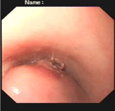
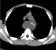
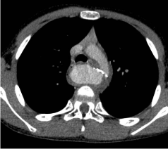
An 11-year-old boy presented with mid-upper retrosternal and abdominal pain for 5 days with haematemesis twice during the abdominal pain. There was around 600-700ml of haematemesis and the blood was dark in color. Prior to the day of admission, he had an episode of black stool. There was no significant past medical history. On physical examination, he looked pale. There was tenderness over right lower abdomen without rebound tenderness. His laboratory reports showed WBC as 11.3×10⁹/L with 76% of neutrophil and hemoglobin as 86g/L. Taking into consideration of haematemesis and melena as a predominant complaint on this consultation, following endoscopy revealed mucosal bulge of 2.2cm×5cm with ulceration at the centre of bulging mass together with blood clot at 22-25cm distance from incision (Figure 1) and stomach was normal. Non-enhanced and contrast-enhanced CT scan was required to clarify esophageal mass, which revealed proximal descending aortic Pseudoaneurysm due to esophageal foreign body (FB) (Figure 2,3). Immediately, the patient underwent aortic reconstruction after removing aortic Pseudoaneurysm and esophageal perforation repair under general anesthesia. FB identified by CT is an alloying metal which is used to clean the utensils, which was engulfed unnoticingly when he recalled a past history of eating hot spicy dips. The post-operative course was uneventful, and he was free of pain and haematemesis on the followup of 12 months.

Figure 1:

Figure 2:

Figure 3:
Aortoesophageal fistula (AEF) by FB has been often considered to be a fatal condition. Less than 1% of ingested FB cause perforation of the gastrointestinal tract, even rare is an aortic Pseudoaneurysm with the consequence of an Aortoesophageal fistula caused by FB [1]. AEF by FB most often concurs with the infection of the surrounding tissues; however mediastinal infection was not obvious in our case. According to our presumption, thin metal foreign body rarely results in obvious inflammatory reaction, as described in this patient. As reported in the literatures, the formation of the aortic Pseudoaneurysm could be the result of trauma by FB or by infection. When there is delay between ingestion and presentation, the aortic perforation may be mycotic [2], however in our case we didn’t come to know the exact time of ingestion as it was unknowingly ingested by the boy with his meal and was only found after searching for the cause of gastrointestinal tract bleeding. Chiari have ever given an appropriate description for AEF’s symptoms, namely a triad of symptoms including mid thoracic pain, sentinel arterial upper gastrointestinal hemorrhage and exsanguinations after a variable interval of time. These symptoms are more specific in patients with AEF from FBs than from other causes.Endoscopy and contrast-enhanced CT scan would be the optimal diagnostic tool for defining AEF from FBs, including the site of bleeding, FB, associated infection and Pseudoaneurysm of the aorta. Early diagnosis and prompt management should be the mainstay for treating AEF, as this condition could be a life threatening. The definitive treatment is open surgical repair of both esophagus and aorta.
References