Case Report
Austin J Clin Case Rep. 2016; 3(1): 1084.
Chasing the Dragon - Heroin Inhalation in Saudi Arabia: Case Report
Harbi-Hassan Mohammed Al¹*, Nabil A², Moh’d S² and Naif A¹
¹Department of Internal Medicine, King Faisal Specialist Hospital & Research Center, KSA
²Department of Critical Care Medicine, King Faisal Specialist Hospital & Research Center, KSA
*Corresponding author: Harbi-Hassan Mohammed Al, Department of Internal Medicine, King Faisal Specialist Hospital & Research Center, Riyadh, King Faisal Medical City for Southern Regions, KSA
Received: February 01, 2016; Accepted: April 08, 2016; Published: April 11, 2016
Abstract
Heroin abuse associated death may relate to overdose or associated IV drug abuse disease. We report case of young man who is well-known to be a drug abuser came to the emergency department with low Glasgow coma scale and brain Magnetic Resonance Imaging (MRI) showed diffuse hyper-intensity involving white matter, consistent with heroin vapor encephalopathy. Thus it is important to keep this association in mind for physicians when working up a patient with history of drug abuse presenting with decrease level of consciousness and Brain MRI findings of diffuse, symmetrical white matter hyper-intensities to consider diagnosis of heroin inhalation Leukoencephalopathy.
Keywords: Heroin; MRI; Leukoencephalopathy; Drug abuse
Introduction
Substance abuse is one of the epidemic diseases, its impact extended to family and society at large not on individual only. In Saudi Arabia Abusing drugs and alcohol are consider forbidden by Islam. Addictive behaviors are socially unacceptable In spite of these prohibitions; some people drink, use drugs and become addicted to these substances. Using of illicit drugs in Saudi Arabia usually started with tobacco smoking around age of adolescent and amphetamine noticed to be the first drug to be used after tobacco smoking Among Saudi patients in addiction treatment centers [1]. Saudi government has recognize these as a public health problem and many addiction treatment centers have been established for the treatment of substance abuse.
In developed countries Heroin abuse was a popular drug in last three decade [2]. Heroin abuse -associated death may due to substance abuse itself or related to IV drug abuse disease. Heroin vapor Leukoencephalopathy, was first reported in 1982 from Amsterdam [3]. After that, cases have been reported from time to time in different countries. Heroin abuse is commonly taken by injection; it may also be taken by inhalation of heated vapors.
Case Presentation
A 22-year-old Saudi male admitted to critical care unit after was found collapsed in his room with some pills around as stated by his father. The patient was well-known to be a drug abuser for around 1 year especially with heroin inhalation, with no prior medical history. He was last time seen well two hour prior to his collapse.
Upon arrival to our emergency department Glasgow coma score was 6/15, later patient developed seizure attack, followed by vomiting and aspiration of gastric content and subsequently patient desaturated and urgent intubation was done. Patient received IV phenytoin. And noted to have fever of 38oC but was hemodynamically stable.
Neurological examination revealed patient to be comatose, unresponsive to verbal stimuli. His pupils were both mid dilated and sluggishly reactive, corneal reflexes were intact, with good gag reflex. The face was symmetric. Patient had tonic posturing extending both arm. The Motor examination revealed axial myoclonus involving the neck flexors, the pectoral muscles and the abdominal musculature. Limbs withdrew symmetrically on painful stimuli. Deep Tendon reflexes were present throughout all joints, bilateral Upper going Babinski signs were present. General examination revealed tachypnea with clear lungs. No cardiac murmurs and normal abdominal exam.
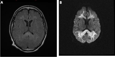
Electroencephalography (EEG) showed abnormal activity consisting of posterior dominant rhythm 3-4 Hz polymorphic delta wave, low amplitude seen at the posterior head region. Continuous generalized slow activity mixture of polymorphic delta wave 2-3 Hz intermixed with 5-6 Hz beta waves, low to moderate amplitude. Intermittent burst of generalized slow activity 2-3 Hz moderate amplitude. In general these findings were suggestive of severe cortical dysfunction and nonspecific encephalopathy. Brain Magnetic Resonance Imaging (MRI) showed brain confluent T2-weighted- Fluid-Attenuated Inversion Recovery (FLAIR) hyper-intensity involving the periventricular, deep, and subcortical white matter, including the corpus callosum with restricted diffusion consistent with acute leukoencephalopathy, a pattern is suggestive of severe heroin vapor encephalopathy Figure 1. Lumbar puncture showed entirely normal cerebrospinal fluid parameter. Chest radiography revealed right upper lobe consolidation.

    Figure 1:  MRI Brain: Diffusion Weighted Image (A) fluid attenuated inversion
recovery FLAIR (B): At the level of centrum semiovale and lateral ventricles,
shows diffuse, symmetrical hyperintensities of the deep and subcortical white
matter, and the corpus callosum.
Urine drug screen was negative for cocaine metabolites and other toxic agents were negative. The Toxicology department analyzed the pills that were found around the patient and showed a combination of dextromethorphan plus heroin.
Over the subsequent 3 weeks. He underwent tracheotomy then weaned off from ventilator and patient became slowly responsive to stimulation in matter of opening eye responding to verbal stimuli until had been discharge from critical care unit to regular ward. Was partially recover having but having spastic limbs with GCS 10 out of 15. And he received antioxidant therapy in form ubiquinone (Coenzyme Q10) in combination with vitamin E with care of multidisciplinary teams include physical therapy, occupational therapy, speech therapy and neurological team.
Discussion
Mortality rate in Heroin leukoencephalopathy reached up to 23% [3]. And has devastating consequences. Heroin was a popular drug in the late 1960s in western country [2]. Among Saudi patients in addiction treatment centers, recorded commonly abused substances were heroin ranging from (6.6–83.6%), amphetamine (4–70.7 %), alcohol (9–70.3%) and cannabis (1–60%). But in last few years there was an increase in the use of cannabis and amphetamine and decrease in the use of heroin and volatile substances [1]. Some of Heroin abuser heats the powder on aluminum foil and inhale the smoke instate of take it as injection form. This practice is known as “chasing the dragon, “Chinsing” or “Chinese blowing” lead to Heroin vapor leukoencephalopathy condition. The mechanism of neurologic injury related to heroin inhalation is unknown. Despite potential mitochondrial dysfunction could have role in the pathogenesis , based on mitochondrial changes on samples from brain biopsy [3]. As well the raise lactate in white matter that described in first American patients with this syndrome on magnetic resonance spectroscopy and by the clinical improvement that occurred with oral coenzyme Q that has been reported in a few patients [4].
The largest report in the literature regarding Heroin vapor leukoencephalopathy was cohort study of 47 heroin vapor inhalers from the Netherlands the including autopsy for 10 of them, toxicological study of heroin samples, investigation of unaffected heroin addicts and analysis of the effects of heroin vapor in animal models, failed to find a toxicological cause of the leukoencephalopathy [3]. The autopsies discovered severe changes in the white matter, termed vaculoating myelinopathy [3].
In all the heroin cases described in the literature the neuropathologic findings changes in the white matte are consistent with our findings in MRI showing hyperintensity involving the periventricular, deep and subcortical white matter, including the corpus callosum. Which is considered a highly specific finding. Our extensive wide literature review revealed no reported other toxic substance encephalopathy causing similar brain destruction. Thus it is important to keep this association in mind for emergency physicians, internists, and neurologists to consider probable relation when working up a patient with history of drug abuse presenting with acute or subacute onset of neurologic manifestation and Brain MRI findings of diffuse, symmetrical white matter hyper-intensities to consider diagnosis of heroin inhalation Leukoencephalopathy.
References
- Sweileh WM, Zyoud SH, Al-Jabi SW, Sawalha AF. Substance use disorders in Arab countries: research activity and bibliometric analysis. Subst Abuse Treat Prev Policy. 2014; 9: 33.
- DuPont RL. Profile of a heroin-addiction epidemic. N Engl J Med.1971; 285:320–324.
- Wolters EC, van Wijngaarden GK, Stam FC, Rengelink H, Lousberg RJ, Schipper ME, et al. Leucoencephalopathy after inhaling “heroin” pyrolysate. Lancet (London, England). 1982; 2:1233–1237.
- Hedley-Whyte ET, Kriegstein AR, Shungu DC, Millar WS, Armitage BA, Brust JC, et al. Leukoencephalopathy and raised brain lactate from heroin vapor inhalation. Neurology. 2000; 54: 2027–2028.