
Case Report
Austin J Clin Case Rep. 2016; 3(1): 1086.
Prenatal Diagnosis of Idiopathic Infantile Arterial Calcification: A Lethal Heritable Condition
Gowda M*, Papa D and Sagili H
Department of Obstetrics and Gynecology, JIPMER, India
*Corresponding author: Mamatha Gowda, Department of Obstetrics and Gynecology, Medical Genetics Unit, JIPMER, Dhanvantri Nagar, Pondicherry-605006, India
Received: January 05, 2016; Accepted: April 04, 2016; Published: April 06, 2016
Abstract
Idiopathic infantile arterial calcification is a rare disorder which is generally fatal and characterized by extensive calcification of large and medium sized arteries. The diagnosis is usually made at autopsy or in the neonatal period, when there is cardiac failure. Prenatal diagnosis is possible in the latter half of pregnancy when there is hyperechoic vessel walls, hypertrophied ventricular musculature and nonimmune fetal hydrops. There are only few cases reported to have been diagnosed antenatally. The inheritance is autosomal recessive and most of the affected individuals have mutations in ENPP1 and ABCC6 genes. Molecular genetic tests and genetic counselling should be offered to provide early prenatal diagnosis in subsequent pregnancies.
Keywords: Idiopathic infantile arterial calcification; Generalized arterial calcification of infancy; Arterial calcification
Introduction
Generalized arterial calcification of infancy (GACI), also known as idiopathic infantile arterial calcification (IIAC) is a rare disease with about 180 reported cases [1]. Prenatal diagnosis has been reported in fewer than 10 cases and represent a severe form with worse prognosis [2]. The case reported here presented with cardiomegaly and pericardial effusion at 36 weeks of gestation. The hyper-echogenicity of large vessels may be missed if one is not aware of the condition. As the disease is heritable in an autosomal recessive manner it is important to make the diagnosis in the proband. Parents should be offered genetic counselling, molecular genetic tests of proband and prenatal diagnosis in subsequent pregnancies. Prenatal diagnosis by ultrasound is usually made in the third trimester and the condition is usually not evident in early pregnancy. Prenatal diagnosis by molecular genetic test is possible early in gestation.
Case Presentation
A multigravida with previous two normal issues was referred at 36 weeks of gestation, as the screening ultrasound revealed cardiomegaly. The patient had received regular antenatal care and an anomaly scan done at 19 weeks of gestation was reported to be normal. There was history of second degree consanguinity. Targeted ultrasonography revealed moderate cardiomegaly and pericardial effusion in the fetus (Figure 1). There were no structural abnormality of the heart on fetal ECHO. The origin of great vessels, the interventricular and interatrial septae and the valves were normal. The venous connection to the atrial chambers were normal. The fetus was noted to have abnormally echogenic walls of great arteries and on closer look, the aortic and pulmonary vessel walls appeared calcified with narrowing of abdominal aorta. The iliac arteries were almost obliterated at their origin (Figure 2). The coronaries were also calcified. A diagnosis of idiopathic arterial calcification was made and patient was recalled the next day.
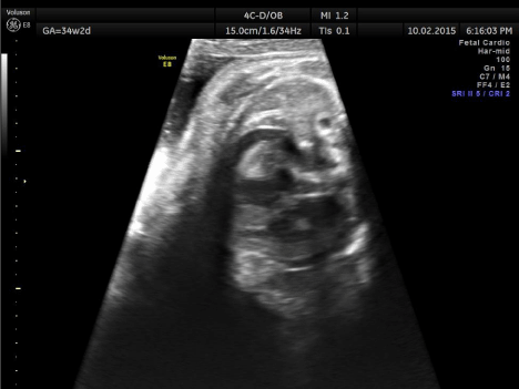
Figure 1: Cardiomegaly, hypertrophied ventricles and pericardial effusion.
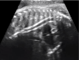
    Figure 2:  Gowda M, Papa D and Sagili H. Prenatal Diagnosis of Idiopathic Infantile Arterial Calcification: A Lethal
Heritable Condition. Austin J Clin Case Rep. 2016; 3(1): 1086.
The mother presented with intra-uterine demise of the fetus, a day after the diagnosis, and delivered a fresh stillborn male baby weighing 3000g. There were no associated malformations on external examination of the baby. Parents had declined fetal autopsy and further genetic tests as they were not planning another pregnancy.
Discussion
Infantile arterial calcification was first described in 1901 by Bryant and White [3]. It is a rare disease characterized by extensive calcification of medium and large arteries. There is calcium hydroxyapatite deposition in the internal elastic lamina, disruption of elastic fibers and extensive intimal fibrous proliferation [4].
It is mostly diagnosed in the neonatal period or early infancy and presents with rapidly progressive heart failure and refractory hypertension. The condition is nearly always fatal. Prenatal diagnosis is mostly made at the beginning of third trimester. The fetus may have nonimmune hydrops fetalis, polyhydramnios, hypertrophic cardiomyopathy, pericardial effusion, highly echogenic great vessel walls with narrowing of aorta at its bifurcation into common iliac arteries.
Clinical Presentation
The spectrum of the disease varies from fetal demise in later pregnancy to refractory hypertension and rapidly progressive ischemic heart failure within few months of birth. Coronary arteries are almost always involved and 85% of affected infants die within 6 months due to myocardial ischemia/infarction [4].
Other less common clinical presentations are, joint swelling with periarticular calcification, gangrene of extremities, visceral infarction, seizures, progressive hepatic failure and cerebral atrophy [5,6]. More severe cases of non-immune hydrops fetalis results from hypertension, ventricular hypertrophy and cardiac failure due to dystrophic calcification [2,7].
A case series by Nasrallah et al., described three fetuses with GACI, one of which was diagnosed by 23 weeks. All three cases had an echogenic intracardiac focus at 20 weeks’ gestation. The presence of echogenic intracardiac focus was suggested to be an early sign of GACI in patients with family history.
There are other reports of prenatally detected cases of GACI. The condition may also present as in utero meconium peritonitis due to mesenteric ischemia [8] Wax et al. reported a case that presented with hepatic vascular calcification at 18th weeks of gestation [9].
Diagnosis
Most cases of IIAC are diagnosed at autopsyor during first months of infancy [1]. Prenatal diagnosis is extremely rare with about 10 reported cases [2,7,10]. The gold standard for diagnosis of GACI is arterial biopsy [3]. A combination of CT, MRI, plain X-ray and sonography are alternatives to this invasive technique [11]. Prenatal diagnosis is by sonographic examination. The walls of large vessels appear hyperechoic, ventricles are dilated and there may be hydrops fetalis [3]. As the vascular calcification may not be visible until late gestation or postnatally, the diagnosis cannot be easily made in early pregnancy.
Genetic Basis
GACI is reported to be caused by mutations in ENPP1 gene and ABCC6 gene. ENPP1 gene is located on Chromosome 6q22 and mutation of the gene inactivates ecto-nucleotide pyrophosphatase/ phosphodiesterase 1. Pyrophosphatase/phosphodiesterase 1 is involved in generation of the inorganic pyrophosphate which is a physiologic inhibitor of calcium deposition [12]. Rutsch et al. identified 40 different homozygous or compound heterozygous mutations in 75% of patients with GACI [13]. Nitschke et al. identified 13 different mutations in ABCC6 gene in 28 GACI patients [14].
IIAC is inherited as an autosomal recessive disorder with a 25% recurrence risk in future pregnancies ]15]. Genetic counselling is very important and molecular analysis of ENPP1 and ABCC6 genes should be offered to the family. If disease-causing mutations have been identified in the index case, early prenatal diagnosis by mutation analysis is possible [16].
Treatment
GACI is generally lethal and various treatment modalities such as estrogens, steroids and bisphosphonates have been described [1]. There are few reports of response to treatment with biphosphonates. As pyrophosphate derivatives, Biphosphonates may prevent or reverse abnormal calcium deposition in patients with abnormal serum calcium levels. However, the side effects and doubtful efficacy outweigh its benefits as therapy for this condition [16]. The role of intrauterine administration of biphosphonate is also not clear. Medical treatment of cardiovascular complications is usually unsuccessful [17]. Spontaneous resolution of calcification has occasionally been reported but the long-term prognosis in survivors is not described [18].
Conclusion
GACI is a rare, fatal disease of extensive arterial calcification usually diagnosed in later pregnancy or postnatal period. The disease can present as fetal hydrops during pregnancy or with cardiac failure postnatally. When there is a family history, serial sonographic examination for signs of calcification may allow early diagnosis. Genetic counselling and molecular analysis should be offered in index cases to provide early prenatal diagnosis in subsequent conceptions. The disease is generally lethal and the role of medical therapy needs to be explored.
References
- Rosenbaum DM, Blumhagen JD. Sonographic recognition of idiopathic arterial calci?cation of infancy. AJR Am J Roentgenol; 1986; 146: 249–250.
- Nasrallah FK, Baho H, Sallout A. Prenatal diagnosis of idiopathic infantile arterial calci?cation with hydrops fetalis. Ultrasound Obstet Gynecol; 2009; 34: 601-604.
- Bryant JH, White WA. A case of calcification of the arteries and obliterative endarteritis associated with hydronephrosis in a child aged 6 months. Guys Hosp Rep; 1901; 55: 17–28.
- Farquhar J, Makhseed N, Sargent M. Idiopathic infantile arterial calci?cation and persistent pulmonary hypertension. Am J Perinat; 2005; 22: 121-125
- Van der Sluis IM, Boot AM, Vernooij M. Idiopathic infantile arterial calci?cation: clinical presentation, therapy and long-term follow up. Eur J Pediatr; 2006; 165: 590-593.
- Whitehall J, Smith M, Altamirano L. Idiopathic infantile arterial calci?cation: sonographic ?ndings. J Clin Ultrasound; 2003; 31: 497-501.
- Nagar AM, Hanchate V, Tandon A. Antenatal detection of idiopathic arterial calci?cation with hydrops fetalis. J Ultrasound Med; 2003; 22: 653-659.
- Sawyer T, Stacey M, Mulreany M. Generalized arterial calci?cation of infancy associated with meconium peritonitis: a case report and review of the literature. Am J Perinat; 2009; 26: 711-716
- Wax JR, Balckstone J, Pinette MG. Hepatic vascular calci?cation: an early second trimester sonographic feature of idiopathic infantile arterial calcinosis. Am J Obstet Gynecol; 2001; 185: 1267-1268.
- Azancot A, Diehl R, Dorgeret S, Sebag G. Isolated pericardial effusion in the human fetus: a report of three cases. Prenat Diagn; 2003; 23: 193–197.
- Greenberg SB, Gibson J. New findings in idiopathic arterial calcification of infancy detected by MDCT. AJR Am J Roentgenol; 2005; 185: 530–532.
- Rutsch F, Ruf N, Vaingankar S. Mutations in ENPP1 are associated with ‘idio- pathic’ infantile arterial calcification. Nat Genet; 2003; 34: 379–381.
- Rutsch F, Boyer P, Nitschke Y. Hypophosphatemia, hyperphosphaturia and bisphosphonate treatment are associated with survival beyond infancy in generalized arterial calci?cation of infancy. Circ Cardiovasc Genet; 2008; 1: 133-140
- Nitschke Y, Baujat G, Botschen U. Generalized arterial calci?cation of infancy and pseudoxanthoma elasticum can be caused by mutations in either ENPP1 or ABCC6. Am J Hum Genet; 2012; 90: 25-39.
- Samon LM, Ash KM, Murdison KA. Aorto-pulmonary calci?cation: an unusual manifestation of idiopathic calci?cation of infancy evident antenatally. Obstet Gynecol; 1995; 85: 863–865.
- Kalal IG, Seetha D, Panda A. Molecular diagnosis of generalized arterial calci?cation of infancy (GACI). J Cardiovasc Dis Res; 2012; 3: 150-154.
- Eronen M, Pohjavuori M, Heikkila P. Fatal outcome of 2 siblings with idiopathic arterial calcification of infancy diagnosed in utero. Pediatr Cardiol; 2001; 22:167–169.
- Marrott PK, Newcombe KD, Becroft DM, Freidlander DH. Idiopathic infantile arterial calcification of infancy with survival to adult life. Pediatr Cardiol; 1984; 5:119–122.