
Clinical Image
Austin J Clin Case Rep. 2016; 3(1): 1087.
Right Coronary Artery from the Non-Coronary Cusp
Miller RJH, Ramlal R and Mylonas I*
Department of Cardiac Sciences, University of Calgary, Canada
*Corresponding author: Mylonas I, Department of Cardiac Sciences, University of Calgary, 3rd floor, 8031 Ave NE, Calgary, AB T2E 0C2, Canada
Received: March 02, 2016; Accepted: April 15, 2016; Published: April 18, 2016
Clinical Image
A 47-year-old woman was evaluated for atypical chest pain with a prospectively gated, cardiac CT coronary angiogram (CTA) after previous perfusion imaging had shown coronary calcium without evidence of ischemia. The CTA demonstrated that the right coronary artery (RCA) arose from the non-coronary cusp, as shown in Figure 1 and 2. There was no ostial stenosis and the course of the artery appeared free from compression, therefore this was believed to be a low-risk anomaly. There was no evidence of coronary artery disease and no other significant findings. Coronary anomalies are present in 1-5% of the general population. CTA accurately identifies coronary anomalies and provides an assessment of their potential significance. This particular coronary anomaly has only been described once previously, but could readily be classified as low-risk based on its appearance and benign vascular course.
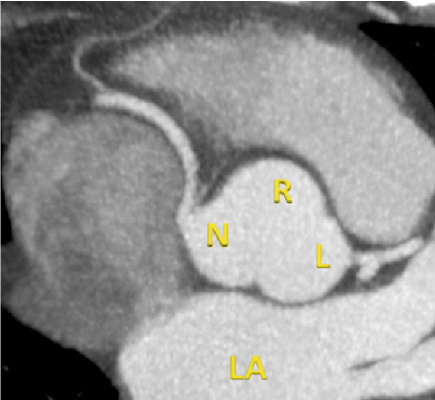
Figure 1:
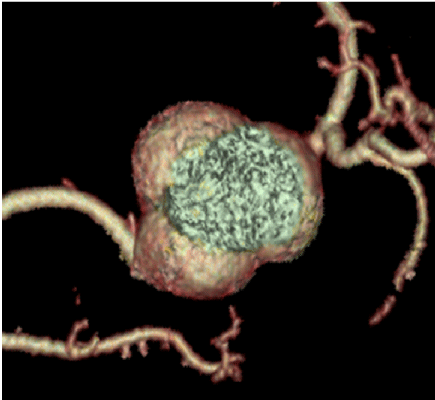
Figure 2: