
Case Report
Austin J Clin Case Rep. 2016; 3(2): 1092.
Spine Metastasis of Conjunctival Melanoma: A Case Report
Xue Y¹, Ma J2#, Liu X¹, Li H¹ and Fan H¹*
¹Department of Orthopaedic Surgery, Fourth Military Medical University, China
²State Key Laboratory of Cancer Biology & Institution of Digestive Disease, Fourth Military Medical University, China
#Author contributed to the work equally and should be regarded as co-first author
*Corresponding author: Hongbin Fan, Department of Orthopaedic Surgery, Xi-Jing Hospital, Fourth Military Medical University, Xi’an, 710032, China
Received: May 26, 2016; Accepted: July 05, 2016; Published: July 12, 2016
Abstract
Purpose: Conjunctival melanoma (CM) is rare in ocular melanomas, which often develop systemic metastasis. As the latent event of melanoma progression, bone metastasis is not common. The tumor with high invasion may result in the bone structure destruction, fracture occurrence, and motor dysfunction. Till now, there are few reports on bone metastasis of conjunctival melanoma.
Methods: A 53-year-old woman who had a backache and ribs pain for 6 months was diagnosed of multiple melanoma metastasis of spine. MRI indicated the metastasis tumor of T11-T12 spine and pathological fracture. The bone scan found disseminated mass radioactivity concentration points in spine, ribs, and limbs. Decompressive laminotomy was performed in T11-T12 to relieve pain and internal fixation in T9-L2 was taken to enhance the stability of spine.
Results: The pathology indicated that the metastasis of malignant melanoma in thoracic vertebrate. Immunohistochemistry staining revealed: HMB-45(+), Pan-mcl(+), S-100(+), CD117(+), NF(-), CD34(-), AE1/AE3(-), and Ki-67 incremental index (40%). The gene mutation report indicated that multiple mutations of C-KIT (N655D mutation), PDGFRA (L641P mutation), and NRAS (E31K mutation) gene. The gene target therapy (imatinib mesylate) was applied and showed satisfactory results.
Conclusion: This report described the incidence of bone metastasis of CM, which had a low survival rate and a poor prognosis. For timely diagnosis and effectively treatment, more detailed examinations should be considered to confirm whether there exists bone metastasis.
Keywords: Conjunctival melanoma; Spine metastasis; Pathological fracture
Introduction
Although conjunctival melanoma (CM) is an uncommon malignancy with the incidence of 0.2 cases per million, it is a sightand life-threatening condition [1]. It represents approximately 1-2% of ocular melanoma and 1.6% of all non-cutaneous melanomas [2,3]. Despite the therapeutic advances in the treatment of CM, local recurrence rates are as high as 62% and mortality ranging from 18% to 44% [4]. Metastases can occur via lymphatics to regional lymph nodes and haematogenously to other parts of the body. Till now, there are few reports on bone metastasis of CM.
Case Presentation
A 53-year-old woman who had suffered severe back and ribs pain for 6 months was referred to our department in 2013. In 2000, she found a linear dark deposit in her left ocular conjunctiva diagnosed of “melanin deposit”. It developed and became a patch like deposit in 2008 (Figure 1A). Physical examination showed obvious kyphosis deformity. The tenderness sites could be found in T11-L2. The muscle strength of both lower limbs was normal and no pathologic reflex sign was observed. The plain film indicated the radiolucent lesions in T11 vertebrae and left 3 to 7 ribs. The sagittal T2-weighted MRI image indicated the multiple spine metastasis and pathological compression fracture in T11 (Figure 1B). The axial MRI image showed low signal lesion invaded nearly all the vertebral body and pedicles (Figure 1C). The PET-CT scan showed extensive increased uptake in spine, ribs, and limb bones (Figure 2A). CT scan showed a large osteolytic lesion involving nearly the whole vertebrae (Figure 2B). In addition, the level of tumor-related serum biomarkers including Lactate dehydrogenase and CA-199 increased.
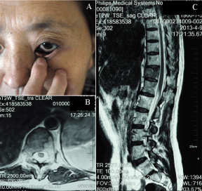
    Figure 1: (A) The photograph illustrated the patient with melanin deposit in
her left ocular conjunctiva; (B) Axial T2-weighted MRI showed low signal area
(tumor lesion) invaded nearly all the vertebral body and pedicles; (C) Sagittal
T2-weighted MRI showing low signal in T11 vertebra with compression
fracture.
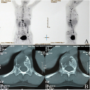
    Figure 2: (A) The PET-CT scan showed extensive increased uptake in spine,
ribs, and limb bones; (B) CT scan showed a large osteolytic lesion involving
nearly the whole vertebrae.
In order to relieve the pain, decompressive laminectomy was performed in T10-T11 level. In operation, the disseminated spotted dark lesions in the vertebral laminae could be observed (Figure 3A). Internal fixation with pedicle screws in T8-L1 level was done to increase the stability of spine (Figure 3B & 3C).
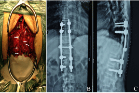
    Figure 3: (A) The photograph illustrated the disseminated spotted dark tumor
tissue in the vertebral laminae could be observed(arrow); The post-operative
AP and lateral radiographs (B, C) show internal fixation with pedicle screws in
T9-L2 level was done to increase the stability of spine.
The pathology indicated the conjunctival melanoma metastasis of spine. Immunohistochemistry results showed the staining of HMB- 45, Pan-mcl, S-100, and CD117 was positive; the staining of NF, CD34, and AE1/AE3 was negative. The Ki-67 incremental index was 40%.
The gene mutation report found multiple mutations such as the stem-cell factor receptor (C-KIT) (chromosome 13 exons; N655D), the platelet-derived growth factor receptor A (PDGFRA) (chromosome 14 exons; L641Pn) and NRAS (chromosome 1 exons; E31K). Due to the weakness, the patient refused to receive the chemotherapy and radiotherapy. However, according to genes analysis report, she started to take imatinib mesylate and was still alive in the latest follow-up (2.5 years post-operation).
Discussion
Shakur et al. [5] reported a case with ocular melanoma metastasis to the cervical spine, in which C1-C2 were destructed severely by tumors. As the spinal cord was compressed progressively by metastatic tumor tissue, the patient presented severe neurological deficits. Due to poor prognosis, the mainstay of therapy is palliative treatment. Surgical decompression and fixation is indicated to preserve neurological function and spine stability. After series of treatments, this case died 1 year later after diagnosis.
In patients with bone metastasis, pain is the most common symptom. Mechanical instability after bony destruction, nerve roots compression, and/or irritation by tumor caused radicular pain. Motor dysfunction is another common symptom. Uracratia is common when patients have autonomic dysfunction [6]. Sensory dysfunction can combined or only exist in some cases.
In immunohistochemistry staining, S-100 protein (the common marker) and HMB-45 (with high specificity) [7] have clinical significance in diagnosis but no meanings in prognosis. Tumor markers can be positive when bone metastasis occurs but have limited specification to identify the primary tumor site [8]. Some gene mutation can be found in the metastasis of CM including BRAF V600, CDKN1A, RUNX2, MLH1, TIMP2, MGMT, and KIT [9,10]. Imatinib mesylate is a recently developed oral anticancer agent, which selectively inhibit certain cell-surface protein tyrosine kinases including PDGF-R and c-Kit. Activation of c-Kit, often in association with a mutation of the c-kit proto-oncogene, is believed to be present in all cases of gastrointestinal stromal tumor (GIST). Dysregulated PDGF-R function is associated with gliomas, myeloproliferative disorders, melanomas, carcinomas, and sarcomas, including dermatofibrosarcoma protuberans [11]. In this report, imatinib mesylate was used to treat CM and showed satisfactory results.
Conclusion
Low incidence and unpredictable metastasis generate the fact that diagnosis of CM is usually made only after obvious metastasis symptom presentation, which will pose a great challenge to treatment. In recent study, multiple gene mutation might play a role in the incidence of melanoma metastasis. Therefore, target gene therapy can offer a new treatment.
References
- Shildkrot Y, Wilson MW. Conjunctival melanoma: pitfalls and dilemmas in management. Curr Opin Ophthalmol. 2010; 21: 380-386.
- Damato B, Coupland SE. An audit of conjunctival melanoma treatment in Liverpool. Eye (Lond). 2009; 23: 801-809.
- Shields CL. Conjunctival melanoma: risk factors for recurrence, exenteration, metastasis, and death in 150 consecutive patients. Trans Am Ophthalmol Soc. 2000; 98: 471-492.
- Lim LA, Madigan MC, Conway RM. Conjunctival melanoma: a review of conceptual and treatment advances. Clin Ophthalmol. 2013; 6: 521-531.
- Shakur SF, Takagi I, Lukas RV. Ocular melanoma metastasis to the cervical spine. J Clin Neurosci. 2012; 19: 610-611.
- Schiff D. Spinal cord compression. Neurol Clin. 2003; 21: 67–86.
- Glasgow BJ, McCall LC, Foos RY. HMB-45 antibody reactivity in pigmented lesion of the conjunctiva. Am J Ophthalmol. 1990; 109: 696–700.
- Destombe C, Botton E, Le Gal G. Investigations for bone metastasis from an unknown primary. Joint Bone Spine. 2007; 74: 85-89.
- Lake SL, Jmor F, Dopierala J. Multiplex ligation-dependant probe amplification of conjunctival melanoma reveals common BRAF V600E gene mutation and gene copy number changes. Invest Ophthalmol Vis Sci. 2011; 52: 5598–5604.
- Dai B, Cai X, Kong YY. Analysis of KIT expression and gene mutation in humanacral melanoma: with a comparison between primary tumors and corresponding metastases/recurrences. Hum Pathol. 2013; 44: 1472-1478.
- Guilhot F. Indications for imatinib mesylate therapy and clinical management. Oncologist. 2004; 9:271-281.