Case Report
Austin J Clin Case Rep. 2016; 3(3): 1096.
Multiple Myeloma Presenting as Cryoglobulinemia - A Case Report
Narayanan G*, Prabhakaran P and Soman LV
Department of Medical Oncology, Regional Cancer Centre, India
*Corresponding author: Geetha Narayanan, Department of Medical Oncology, Regional Cancer Centre, Trivandrum 695011, Kerala, India
Received: June 01, 2016; Accepted: August 30, 2016; Published: September 09, 2016
Abstract
Cryoglobulinemia is a rare disorder characterized by the presence of abnormal immunoglobulins in the blood that precipitate in the tissues causing inflammation and tissue damage. It often occurs in association with diseases such as autoimmune or infectious diseases. Only few cases of cryoglobulinemia associated with multiple myeloma has been described. We report a 45 year old lady with multiple myeloma whose initial presentation was cryoglobulinema with vasculitic ulcers in legs and gangrene of toes. She had monoclonal gammopathy of IgG kappa. She received chemotherapy with bortezomib, lenalidamide and dexamethasone. Her pain symptoms were controlled and her skin lesions healed. She is alive in remission at 30 months.
Keywords: Cryoglobulinemia; Vasculitis; Multiple myeloma
Abbreviations
CG: Cryoglobulinemia; IFE: Immunofixation; Ig: Immunoglobulins; ISS: International Staging System; MM: Multiple Myeloma; SPE: Serum Protein Electrophoresis
Introduction
Cryoglobulinemia (CG) is a rare disorder characterized by the presence of abnormal immunoglobulins (Ig) in the blood that precipitate in the tissues at low temperatures causing inflammation and tissue damage. CG often occurs in association with diseases such as autoimmune or infectious diseases. It is classified in to 3 major types, type 1 accounts for 10-15% of CG and is associated with hematologic malignancies, type 2 and 3 are associated with autoimmune disorders and chronic infections [1]. CG tends to affect females between the ages 40-60 years of age. Only few cases of CG associated with Multiple Myeloma (MM) has been described. We report a patient with multiple myeloma whose initial presentation was cryoglobulinema and vasculitis.
Case Presentation
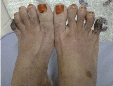
A 45 year old lady presented to us with history of repeated ulcerations in lower limbs since 3 years, numbness and pain since 1 year, and blackish discoloration of little toes on both legs since 1 month. On examination she had multiple ulcers in various stages of healing over both lower limbs, diffuse skin rash similar to livido reticularis and gangrene of little toes on both legs (Figure 1). There was no lymphadenopathy or hepatosplenomegaly. Other systems were normal.

    Figure 1:  Picture showing vasculitic ulcers on the right leg. Picture showing
gangrene of both little toes.
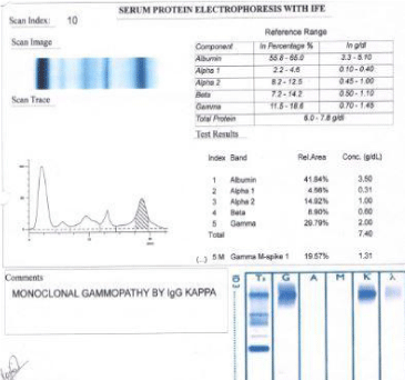
Her haemoglobin was 11.3 gm%, total leucocyte count was 9600/mm3, platelet count was 255000/mm3 and ESR was 88 mm/ hr. Serum creatinine was 2.2 mg/dl and s.calcium was normal. Total protein was 8.1 gm/dl with albumin globulin reversal. Serum Protein Electrophoresis (SPE) showed monoclonal gammopathy, with M protein of 1.4 gm/dl (Figure 2). On quantitative immunoglobulin assay, IgG was 2749 mg/dl, IgA was 139 mg/dl, IgM was 73 mg/dl.

Figure 2: Serum Electrophoresis and Immunofixation of the patient.
The serum free Kappa was 134 mg/L, free lambda was 21 mg/L and K:L ratio was 6.4. Immunofixation Electrophoresis (IFE) showed monoclonal gammopathy of IgG kappa (Figure 2). Her 24 hour urine protein was 235 mg/day, β2 microglobulin was 9.4 mg/L, and urine bence jones protein was negative. She did not have bone lesions. A bone marrow study showed increase in plasma cells. Skin biopsy was suggestive of leucoclastic vasculitis, nerve conduction showed signs of moderate degree of bilateral carpal tunnel syndrome in upper limbs. Cryoglobulin test was positive, rheumatoid factor was absent. She was staged by International Staging System (ISS) as ISS 3.
A diagnosis of multiple myeloma with cryoglobulinemia and vasculitis was made. She was started on chemotherapy with bortezomib, lenalidamide and dexamethasone. Her pain symptoms disappeared after 2 cycles with healing of vasculitic ulcers occurring by 4th cycle. She received 6 cycles of chemotherapy at the end of which her IgG was 686 mg/dl, free kappa was 15.7 mg/dl, s.creatinine 1.3 mg/dl. Her SPE became normal and β2 microglobulin was 3.10 mg/ dl. Her renal functions improved and cryoglobulins disappeared from the blood. Her vasculitic ulcers healed. She refused autologous stem cell transplantation. She has completed 1 year lenalidamide maintenance and is currently in remission at 30 months.
Discussion
The prevalence of clinically significant cryoglobulinemia has been estimated at approximately 1 in 100,000 [2]. Type I CG is composed of monoclonal Ig; type II CG consisting of a combination of polyclonal and monoclonal Ig; and type III CG characterized by a combination of polyclonal Ig [3]. The type I CGs are Igs that precipitate at temperatures below 37°C. They are usually composed of a monoclonal Ig, mainly IgG (IgG1 and IgG3) and occasionally IgM or IgA. Type I cryoglobulins account for 10-15% of total CGs [1]. They are found mostly in patients with low-grade lymphoproliferative disorders, such as Waldenström Macroglobulinemia or MM [4]. About 6-10% of patients with cryoglobulinemia, are diagnosed with myeloma [5]. In a cohort of IgG MM, the frequency of detectable CGs, was 10%, and only a few patients had symptoms related to CG [6].
In general, CGs, regardless of their type, are associated with skin manifestations, but may also cause rheumatologic symptoms that involve the kidneys, the peripheral nerves or central nervous system, the lungs, the myocardium and the gastrointestinal tract [4]. Type I CG classically produces signs related to hyperviscosity or small vessel vasculitis and/or thrombosis: Raynaud phenomenon, digital ischemia, livedo reticularis, and purpura may occur progressing to gangrene in severe cases. Neurologic symptoms of hyperviscosity include blurring or loss of vision, headache, vertigo, sudden deafness, diplopia, ataxia, confusion, and disturbances of consciousness [2].
Cutaneous lesions are unusual during the course of multiple myeloma. In rare cases, multiple myeloma may be associated to skin involvement secondary to amyloidosis, cryoglobulinemia, and POEMS syndrome. Cutaneous manifestations vary from purplish rashes to necrosis and gangrene.
In a cohort of 86 patients with cryoglobulinemia, eight patients had MM, of which 6 had type I CG [3]. In another study on 72 patients with CG, 14 had a lymphoproliferative disorder [7]. Among 913 patients with CG, only four patients had MM [8]. Only 2 patients had a hematologic disease among 31 cases of type I CG [2]. A French retrospective study identified 64 patients with symptomatic type I CG who had either a hematologic malignancy or monoclonal gammapathy [9].
A 62 year old lady with MM of IgG kappa associated with CG and having ulcers, gangrene of ear lobes, fingers and toes is reported. She underwent plasmapharesis and received thalidomide and dexamethasone [10]. Another 62 year old man with MM and IgG kappa presented with severe ulcers in lower extremities, the biopsy showed leucocytoclastic vasculitis and he was positive for cryoglobulins. He received bortezomib and dexamethasone followed by plasma pharesis [11]. A 50 year old man with extensive soft tissue necrosis of fingers, ear lobes and scrotum was diagnosed as MM and hyperviscosity syndrome, he received plasma exchange, thalidomide and dexona [12]. Another 58 year old lady with IgA lambda MM presented with vascular purpura and cutaneous leukocytoclastic vasculitis which resolved after myeloma treatment [13]. A patient presented with gangrene of all fingers and toes, was finally diagnosed and treated as cryoglobulinemic vasculitis due to multiple myeloma [14]. A 61 yr old man with IgG lambda and IgA kappa myeloma presented with necrotizing skin ulcers, treated with VAD and bortezomib progressed and died [5]. In a series of 7 pts with type 1 CG associated with MM, the median age was 53 years, with male preponderance, were mostly IgG monoclonal and ISS stage 1, and manifested as cryoglobulin symptoms like skin lesions, rheumatologic symptoms, neurologic abnormalities, renal defects [15]. Our patient also had a similar presentation.
The management and prognosis of type I CG in patients with MM are poorly defined. In the French study treatment included glucocorticoids, plasma exchange, alkylating agents, rituximab, and chemotherapy, the ten-year survival was 87%, with poorer survival rates in patients with hematologic malignancy [9]. For severe type I CG, plasmapharesis at the onset and specific MM treatment like bortezomib and lenalidamide at the earliy stage is suggested to avoid CG relapse [15]. As most patients with MM-related type I CG are under 65 years at diagnosis, a therapeutic strategy that is similar to that of MM should be adopted, including bortezomib, dexamethasone, and lenalidomide [15]. Our patient was also treated with bortezomib containing chemotherapy with very good response.
In summary, type I CG associated with MM is only rarely reported. Treatment recommendations include plasmapheresis and treatment of the underlying myeloma. A diagnostic delay can result in severe mutilation and multiple organ damage.
References
- Tedeschi A, Barate C, Minola E, Morra E. Cryoglobulinemia. Blood Rev. 2007; 21: 183-200.
- Trejo O, Ramos-Casals M, García-Carrasco M, Yague J, Jimenez S, de la Red G, et al. Cryoglobulinemia: study of etiologic factors and clinical and immunologic features in 443 patients from a single center. Medicine (Baltimore). 2001; 80: 252-262.
- Brouet JC, Clauvel JP, Danon F, Klein M, SeligmannM. Biologic and clinical significance of cryoglobulins. A report of 86 cases. Am J Med. 1974; 57: 775-788.
- Rieu V, Cohen P, André MH, Mouthon L, Godmer P, Jarrousse B, et al. Characteristics and outcome of 49 patients with symptomatic cryoglobulinaemia. Rheumatology. 2002; 41: 290-300.
- Ninomiya S, Fukuno K, Kanemura N, Goto N, Kasahara S, Yamada T, et al. IgG type multiple myeloma and concurrent IgA type monoclonal gammopathy of undetermined significance complicated by necrotizing skin ulcers due to type I cryoglobulinemia. J Clin Exp Hematop. 2010; 50: 71-74.
- Dispenzieri A. Symptomatic cryoglobulinemia. Curr Treat Options Oncol. 2000; 2: 105-118.
- Cohen SJ, Pittelkow MR, Su WP. Cutaneous manifestations of cryoglobulinemia: clinical and histopathological study of seventy-two patients. J Am Acad Dermatol. 1991; 25: 21-27.
- Monti G, Galli M, Invernizzi F, Pioltelli P, Saccardo F, Monteverda A, et al. Cryoglobulinaemias: a multicentre study of the early clinical and laboratory manifestations of primary and secondary disease. GISC. Italian Group for the Study of Cryoglobulinaemias. QJM 1995; 88: 115-126.
- Terrier B, Karras A, Kahn JE, Le Guenno G, Marie I, Benarous L, et al. The spectrum of type I cryoglobulinemia vasculitis: new insights based on 64 cases. Medicine (Baltimore). 2013; 92: 61-68.
- Ryu H, Park B, Moon JY, Lee MW, Choi YS, Song IK, et al. Gangrenous Cryoglobulinemic Vasculitis in a Patient with Multiple Myeloma. Korean J Med. 2013; 85: 634-638.
- Jiménez-Encarnación E, García-Pallas MV, Vilá LM. Severe leg ulcers in a multiple myeloma patient with cryoglobulinemic vasculitis. P R Health Sci J. 2012; 31: 71
- Medic MG, Brajkovic AV, Bosnic D, Gornik I, Babel J, Gašparovic V. Multiple myeloma presenting with lower extremity gangrene and hyperviscosity syndrome. Signa Vitae. 2014; 9: 99-101.
- Peterlin P, Ponge T, Blin N, Moreau P, Hamidou M, Agard C. Paraneoplastic cutaneous leukocytoclastic vasculitis disclosing multiple myeloma: a case report. Clin Lymphoma Myeloma Leuk. 2011; 11: 373-374.
- Vacula I, Ambrózy E, Makovník M, Stvrtina S, Babál P, Stvrtinová V. Cryoglobulinemia manifested by gangraene of almost all fingers and toes. Int Angiol. 2010; 29: 560-564.
- Payet J, Livartowski J, Kavian N, Chandesris O, Dupin N, Wallet N, et al. Type I cryoglobulinemia in multiple myeloma, a rare entity: analysis of clinical and biological characteristics of seven cases and review of the literature. Leuk Lymphoma. 2013; 54: 767-777.