Case Report
Austin J Clin Case Rep. 2016; 3(3): 1097.
An Uncommon Presentation of Takotsubo Cardiomyopathy
Chaudhary R¹*, Waqas A¹ and Stoeckel J²
¹Department of Internal Medicine, James J Peters VA Medical Center, USA
²Department of Pulmonary and Critical Care, James J Peters VA Medical Center, USA
*Corresponding author: Radhika Chaudhary, Department of Internal Medicine, James J Peters VA Medical Center, USA
Received: June 27, 2016; Accepted: September 07, 2016; Published: September 10, 2016
Abstract
Chest pain is a common reason for admission to the hospital. Amongst the cardiac causes of chest pain acute coronary syndrome continues to be amongst the most common causes. However the other uncommon causes of chest pain should be considered in the young and the middle aged patients presenting with chest pain or in patients with atypical presentations or ECHO features. We present a case report of a young patient presenting with cardiac arrest after a witnessed arrhythmia.
Keywords: Chest pain; Takotsubo cardiomyopathy; takotsubo cardiomyopathy; Stress induced cardiomyopathy; Reversible left ventricular ballooning syndrome
Case Report
A 49 years old female with a past medical history of depression, hypothyroidism, alcoholism, vitamin B12 deficiency and previous gastric bypass procedure six years ago was brought to the ER for altered mental status. She was visiting her friends from another state when she developed strange thoughts and suicidal ideation. She was brought to the psychiatry ER where she was found to be initially hemodynamically stable but subsequently developed generalized tonic clonic seizure. Rapid response was activated and she was found to be in ventricular fibrillation. After cardioversion she returned to sinus rhythm with a heart rate of 104 and blood pressure 140/80. She was intubated and taken to the ICU. She did not have any history of seizure disorder but had eclampsia during her pregnancy. A non contrast CT head was done after initial stabilization and loss of grey white matter differentiation was noted in the left high frontal and parietal area with bilateral basal ganglia suggestive of a diffuse axonal injury.
On day one, she was extubated and her vitals were stable. Her troponin I and CKMB levels were serially monitored which showed a drop after an initial increase (Peak CKMB 335 and peak Troponin I at 0.33 at 4 and 8 hours respectively, after admission). Her EKG was suggestive of QTc prolongation and on 2 D ECHO she was found to have mild left ventricular systolic dysfunction with ejection fraction of 44% (biplane method of disks). There was akinesia of the mid anterior, mid anteroseptal, mid inferior and mid inferolateral wall(s). Doppler parameters were consistent with abnormal left ventricular relaxation (grade 1 diastolic dysfunction). Right ventricle was mildly dilated. Systolic function was mildly reduced.
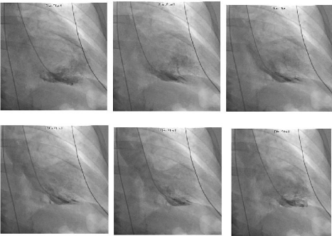
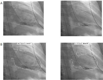
On day two of her admission she underwent a cardiac catheterization. Her coronary arteries were found to be normal but she was found to have mid myocardial akinesia with apical and basal hyperkinesias (Figure 1 & 2). Left ventricular ejection fraction was found to be 40-45%, with left ventricular end diastolic pressure of 17 mm of Hg, suggestive of inverted variant of Takotsubo cardiomyopathy.

    Figure 1:  Sequential images of the ventricle during systole on cardiac
catheterization. The arrow points to the area of mid ventricular dilatation.

    Figure 2:  Cardiac Catheterization images showing the inverted Takotsubo
cardiomyopathy.
A. Cardiac catheterization images showing mid segment ventricular dilatation
with preserved apical function.
B. Delineation of the cardiac borders seen in the image A.
Discussion
Takotsubo cardiomyopathy, also called the reversible left ventricular ballooning syndrome or the stress induced cardiomyopathy [1]. The Mayo Clinic criteria for the diagnosis of SIC recognize this heterogeneity of wall motion abnormalities [2]. In this definition, there is transient hypokinesis, akinesis, or dyskinesis of the LV mid segments with or without apical involvement and the regional wall motion abnormalities must extend beyond a single epicardial coronary distribution. There must be an absence of obstructive Coronary Artery Disease (CAD). This must be accompanied by new EKG abnormalities and modest elevation in cardiac troponin (the peak typically underestimates the degree of wall motion abnormality). Since the original description of this syndrome in Japan (1990) showing apical ballooning with the appearance of a Japanese octopus trap or Tako-tsubo in Japanese [3], the reverse or inverted pattern, a novel variant of this entity has been recognized involving basal and midventricular segments and preserved contractility of apical segment [4-6].
The classical apical Takoysubo cardiomyopathy is believed to be a disease of post menopausal women with a median age of onset of 50 years precipitated by physical or an emotional stress [1,2,6]. Most often the patient presents to the hospital with complaints of chest pain or dyspnea which is often treated as Acute Coronary Syndrome (ACS) until the coronary angiography reveals clean coronary arteries later. Cardiac catheterization of the heart shows the characteristic appearance with a narrow neck and a ballooned lower portion [1,2,6]. The Inverted variant of the Takotsubo differs from the apical Takotsubo with an early age of onset and a more atypical presentation. It has been seen in the setting of a pulmonary embolism [7] and Polytrauma [8]. Though ATSC has been reported to be reversible it can present with severe refractory heart failure requiring balloon pump and in some cases uses of ECHMO has been reported [8].
However inverted Takotsubo cardiomyopathy as a cause of cardiac arrest is less often heard of. We found only one case report of inverted Takotsubo cardiomyopathy, in a patient after cardiac arrest. Most often post cardiac arrest patients have a stunned myocardium resulting in a global decrease in myocardial function. Cvorovic et al. reported a patient who was found to have ITSC on work up of cardiac arrest post anesthesia reversal after a laparotomy in general anesthesia [9].
The exact cause of the TSC is not known but it is believed to be related to expression of adrenergic receptors in the myocardium coupled with stress induced adrenergic surges [1,5-9]. The differential expression of adrenergic receptors in the myocardium in young (basal and mid ventricular) versus the middle aged patients (basal) may explain the differences in the clinical location of the wall motion abdnomality [1]. Post menopausal preponderance has been associated with possible role of estrogen in the prevention of the myocardium from the adrenergic surges in response to stress [7].
Once diagnosed most of the patients have spontaneous recovery with return of the myocardial function to near normal and a low likelihood of recurrence [9]. Some patients may present with cardiogenic shock and require inotropic support which can worsen the cardiac outflow, making the shock worse. Beta blockers and fluid resuscitation remain the main line of treatment. On rare occasions use of IABP (Intra aortic balloon pump) and ECMO (Extracorporeal Membrane oxygenation) has been reported to tide over the acute crisis [6-10].
Conclusion
Takotsubo cardiomyopathy or its inverted variant should be considered as a possible cause of cardiogenic shock. An ECHO finding of apical or mid/basal ventricular dysfunction along the distribution of multiple coronary arteries can be suggestive of the Takotsubo cardiomyopathy or its invert variant. Although it most commonly presents with chest pain or dyspnea, it can present as cardiac arrest. It can occur in multiple clinical settings of physical or emotional stress.
References
- Song BG, Chun WJ, Park YH, Kang GH, Oh J, Lee SC, et al. The clinical characteristics, laboratory parameters, electrocardiographic, and echocardiographic findings of reverse or inverted Takotsubo cardiomyopathy: comparison with mid or apical variant. Clin Cardiol. 2011; 34: 693-699.
- Prasad A, Lerman A, Rihal CS. Apical ballooning syndrome (Tako-Tsubo or stress cardiomyopathy): a mimic of acute myocardial infarction. Am Heart J. 2008; 155: 408-417.
- Kurisu S, Sato H, Kawagoe T, Ishihara M, Shimatani Y, Nishioka K, et al. Tako-tsubo-like left ventricular dysfunction with ST-segment elevation: a novel cardiac syndrome mimicking acute myocardial infarction. Am Heart J. 2002; 143: 448-455.
- Manzanal A, Ruiz L, Madrazo J, Makan M, Perez J. Inverted Takotsubo cardiomyopathy and the fundamental diagnostic role of echocardiography. Tex Heart Inst J. 2013; 40: 56-59.
- Tsuchihashi K, Ueshima K, Uchida T, Oh-mura N, Kimura K, Angina Pectoris-Myocardial Infarction Investigations in Japan, et al. Transient left ventricular apical ballooning without coronary artery stenosis: a novel heart syndrome mimicking acute myocardial infarction. Angina Pectoris- Myocardial Infarction Investigations in Japan. J Am Coll Cardiol. 2001; 38: 11-18.
- Gianni M, Dentali F, Grandi AM, Sumner G, Hiralal R, Lonn E. Apical ballooning syndrome or Takotsubo cardiomyopathy: a systematic review. Eur Heart J. 2006; 27: 1523-1529.
- Lee SH, Kim DH, Jung MS, Lee JW, Nam KM, ChoYS, et al. Inverted-Takotsubo cardiomyopathy in a patient with pulmonary embolism. Korean Circ J. 2013; 43: 834-838.
- Bonacchi M, Vannini A, Harmelin G, Batacchi S. Inverted Takotsubo cardiomyopathy: severe refractory heart failure in a poly- trauma patient saved by emergency extracorporeal life support. Interact Cardiovasc Thorac Surg. 2015; 20: 365-371.
- Cveorovic V, Stankovic I, Panic M, Stipac AV, Zivkovic A, Neskovic AN, et al. Establishing the diagnosis of inverted stress cardiomyopathy in a patient with cardiac arrest during general anesthesia: a potential role of myocardial strain? Echocardiography. 2013; 30: e161-163.
- Sharkey SW, Leeser JR, Maron BJ. Takotsubo (Stress) Cardiomyopathy. Circulation. 2011; 124: e460-462.