Case Report
Austin J Clin Case Rep. 2016; 3(5): 1103.
Choledochal Cyst with Intraductal Papillary Neoplasm of Common Bile Duct
Chao-Wei Lee¹, Cheng-Wei Wang¹, Ming-Chin Yu¹*, Tse-Ching Chen² and Miin-Fu Chen¹
¹Department of Surgery, Chang Gung Memorial Hospital, Linkou, Taiwan
²Department of Pathology, Chang Gung Memorial Hospital, Linkou, Taiwan
*Corresponding author: Ming-Chin Yu, Department of Surgery, Chang Gung Memorial Hospital, Linkou, Taiwan
Received: August 05, 2016; Accepted: October 28, 2016; Published: November 16, 2016
Abstract
Intraductal Papillary Neoplasm (IPN) of Common Bile Duct (CBD) is uncommon, but choledochal cyst coexisting with IPN of CBD is even rare. Due to rarity, the nature of this disease and its treatment outcome are not fully determined yet. We herein report a case of Todani type IV a choledochal cyst with concomitant invasive IPN of CBD.
Case Report
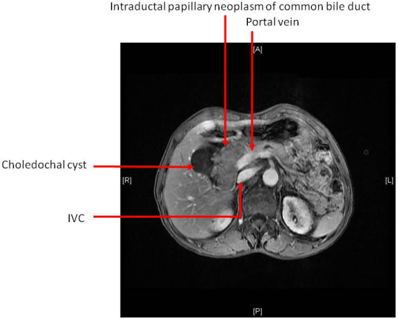
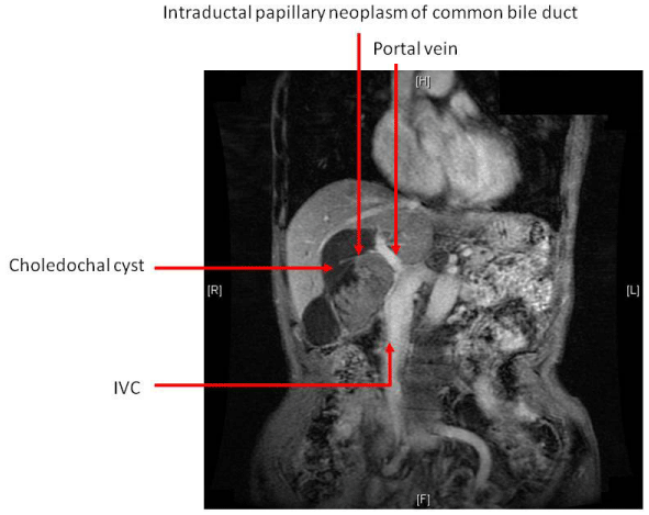
An 88-year-old male farmer suffered from intermittent right upper quadrant abdominal dull pain and fullness sensation for one month. Body weight loss of about 5 kg during this period was also noted. There was no fever, bowel habit change, or tea color urine. The physical examination revealed an 8 x 4 cm ill-defined mass at right upper quadrant of abdomen. No jaundice was found. Tumor markers were within normal range. Both contrast-enhanced computed tomography and Magnetic Resonance Cholangiopancreatography (MRCP) showed markedly dilated Common Bile Duct (CBD) and bilateral Intrahepatic Ducts (IHD) (Figures 1 and 2). Diffuse papillary tumors with slow progressive enhancement were noted within the dilated CBD. Multiple small gallstones were found as well. Todani type IVa choledochal cyst with malignant change was impressed. The patient received total excision of common bile duct, cholecystectomy, and Roux-en-Y hepaticojejunostomy as curative treatment.

Figure 1:

Figure 2:
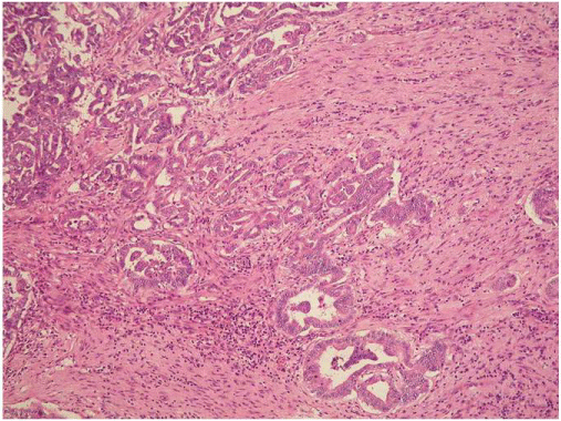
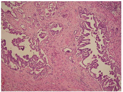
The patient recovered well after the operation and resumed oral intake on postoperative day 5. Pathologic examination revealed a type IVa choledochal cyst (7.8 x 7.2 x 4.0 cm) compacted with a yellow, friable, papillary, and stolid tumor (6.5 x 6.5 x 3.6 cm in dimension). Light microscopy revealed papillary proliferation of ductal cells from the mucosal layer of common bile duct. Under high power field, the ductal glandular epithelium penetrated the lamina propria into fibrous stroma. No obvious atypia was noted. The mitotic index was low (<10/10HPF). The intraductal tumor was composed of mixed pancreatobilliary type, intestinal type, and gastric type (Figures 3 and 4). Intraductal Papillary Neoplasm (IPN) of CBD with an associated invasive adenocarcinoma, pT1N0M0, stage I, was diagnosed. The patient was discharged 2 weeks after the operation and was devoid of complications or tumor recurrence 16 months after the operation.

Figure 3:

Figure 4:
Discussion
Choledochal cyst coexisting with invasive IPN of CBD (IPNCBD) is a rare disease entity. Whenever appropriate, complete resection of CBD could result in satisfactory patient outcome and survival. For radical resection, surgical approach may need liver resection, pancreaticoduodenectomy, or both. Our patient only received complete CBD resection without involvement of liver or pancreas to obtain an adequate oncologic margin and long-term survival. Therefore, organ-preserving surgery should be attempted whenever possible. Although the pancreaticobiliary type of IPN-CBD carries the worst survival prognosis, the nature and behavior of mixed type IPN-CBD with concurrent choledochal cyst has seldom been mentioned and warrants further investigation.