Case Report
Austin J Clin Case Rep. 2016; 3(5): 1104.
An Accessory Spleen Mimicking Left Adrenal Tumor: Tc 99m Labeled Sulfur Colloid Scintigraphy can Avoid Invasive Investigation
Yadav R¹*, Chaudhary S¹, Yadav P² and Kumar A¹
¹Department of Urology, Kidney Transplant & Robotics, Medanta-Medicity, India
²Department of Radiodiagnosis, Medanta-The Medicity, India
*Corresponding author: Yadav Rajiv, Department of Urology, Kidney Transplant and Robotic, Medanta-The Medicity, Gurgaon, India
Received: August 14, 2016; Accepted: October 30, 2016; Published: November 16, 2016
Abstract
We describe a case of accessory spleen mimicking left adrenal tumor. A 48-year-old woman with a history of treatment for locally advanced carcinoma breast was evaluated for left adrenal mass. Due to past history of malignancy, the possibility of solitary metastasis as well as nonfunctional adrenal tumor were considered. Homogenous lesion with the attenuation and enhancement pattern similar to spleen raised the suspicion of an accessory spleen. The finding was confirmed by Sulfur colloid scintigraphy. The article underscores the fact that the urologists should be aware of the possible existence of accessory spleens when left adrenal tumors are suspected on imaging.
Keywords: Adrenal tumor; Accessory spleen; Scintigraphy; Sulfur colloid scan
Introduction
Metastasis to the adrenal is common. Metastatic tumor is the first consideration when a patient with a history of malignancy is found to have adrenal lesion. In the absence of any functional or biochemical abnormalities and equivocal imaging findings, invasive procedures (like biopsy or excision) may be required for characterization of nature of lesion. We present a case of accessory spleen in suprarenal location mimicking adrenal tumor. The presence of accessory spleen was confirmed by Tc 99m labelled Sulfur colloid scintigraphy thus avoiding invasive procedure or surgery.
Case Presentation
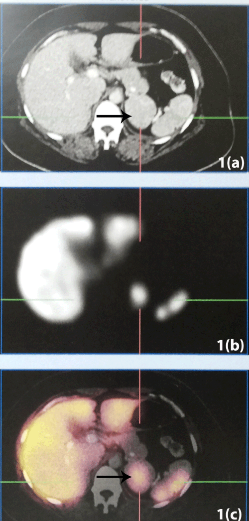
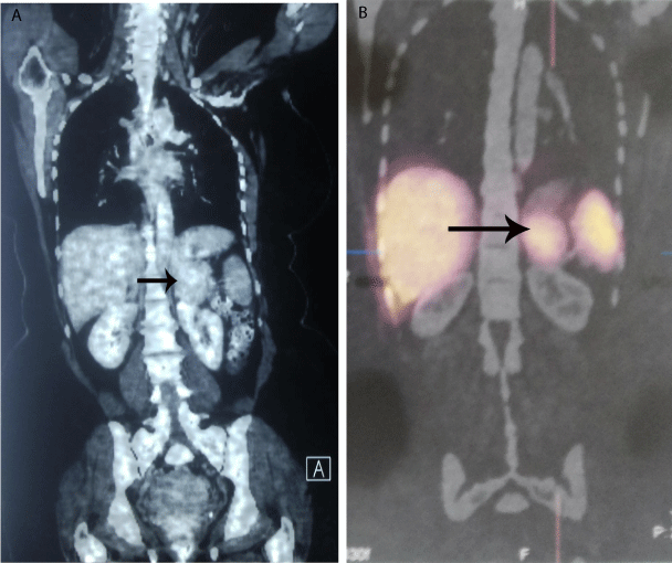
A 48 year old female with a history of treatment for locally advanced carcinoma breast was referred to urology services for evaluation of left supra renal mass. She has had modified radical mastectomy two years ago (Stage T1c, N1a, Mx). Following surgery she had received chemotherapy and Radiotherapy to chest wall and axilla. Left suprarenal mass lesion was first detected on Computed Tomography (CT) in the follow up scan. CT showed a well-defined 4.6 X 4.4 X 4.6 cm, homogenously enhancing mass without any necrosis or calcification in left supra renal area (Figures 1a and 2a). The mass seemed to arise from the lateral limb of left adrenal gland. Lesion showed attenuation value of 40 HU on non-contrast CT and failed to demonstrate significant contrast loss on ‘washout’ study. She is non-hypertensive and non-diabetic. No abnormalities were found on functional and biochemical assessment regarding adrenal lesion.

    Figure 1:  Comparative study showing: 1a) Non-contrast axial CT. 1b) Tc 99m
Sulphur colloid scintigraphy showing accessory splenic tissue. 1c) SPECT
axial image showing accessory spleen showing uptake of labeled colloid in
the lesion. Black arrow marking the accessory spleen.

    Figure 2:  Coronal image: 2a) Contrast CT showing mass lesion in suprarenal
location 2b) SPECT showing uptake of labeled colloid in the accessory
spleen corresponding to the lesion in suprarenal location.
A Positron Emission CT with contrast did not show any other intrabdominal lesion. Left adrenal gland was seen separately, with the lateral limb of adrenal touching the mass. The density and enhancement characteristics of the lesion looked similar to the spleen thus raising the suspicion of accessory splenic tissue. Further to clinch the diagnosis a Tc 99m labeled Sulfur colloid scan was done to differentiate the two. Scintigraphy revealed focally increased tracer
Discussion
Adrenal glands are common site of metastases. All types of common cancers (including breast cancer) can exhibit metastatic deposits in adrenals [1]. Therefore, diagnostic challenge arises when an adrenal incidentaloma is discovered in patients with a known malignancy. Radiographically, metastatic lesions in adrenal also appear similar to benign adenoma on CT (i.e well circumscribed, homogenous and often lack areas of macroscopic necrosis) but lack lipid content [2]. Therefore, in lesions with attenuation value more than 10 HU on non-contrast CT, it may be difficult to differentiate metastatic lesions from lipid poor adenoma or carcinoma. Metastatic lesions as well as carcinomas or atypical adenomas do not show the typical ‘wash out’ pattern seen in adenomas [2]. Therefore, in the absence of any characteristic features of benign adenoma, many such incidentaloma are often subjected to surgery or biopsy especially if the size is >4 cm. Accessory spleen mimicking adrenal tumor is a rare situation. Typically, an accessory spleen is located at the splenic hilum and is less than 3 cm in diameter [3,4]. However, in some situations, its presence at locations other than splenic hilum, may cause clinical problems especially in patients with history of previous malignancy.
Noninvasive methods of detection and confirmation of its presence not only avoid invasive intervention but also allays fear of recurrence of malignancy. As noted in our case accessory spleen is usually round or oval in shape, with CT density similar (or slightly lower) to that of proper spleen thus raising the suspicion of its possibility. In such cases, radionuclide imaging with technetium sulfur colloid may confirm the presence of accessory normal tissue and would therefore avoid invasive investigations or surgery. After intravenous administration, the 99mTc-sulfur colloid accumulates in the tissues of the spleen resulting in their identification as early as after 15- 30 minutes afterwards [5]. Labeled Colloid is phagocytized by the reticuloendothelial cells of the liver, spleen, and bone marrow thus highlighting functional splenic tissue. Further, the examination may be performed with the use of SPECT, thus allowing for evaluation of radiotracer distribution in 3 dimensions. The specificity of this test in identifying accessory spleen is very high however sensitivity may be limited by the size of lesion because accessory spleens of less than 8-10 mm in diameter may cause false-negative results due to poor radiotracer accumulation [6,7]. This case study underscores the fact that the urologists should be aware of the possible existence of accessory spleens when left adrenal tumors are suspected on imaging.
References
- Lenert JT, Barnett CC Jr, Kudelka AP, Sellin RV, Gagel RF, Prieto VG, et al. Evaluation and surgical resection of adrenal masses in patients with a history of extra-adrenal malignancy. Surgery. 2001; 130: 1060-1067.
- Boland GW, Blake MA, Hahn PF, Mayo-Smith WW. Incidental adrenal lesions: principles, techniques, and algorithms for imaging characterization. Radiology. 2008; 249: 756-775.
- Gayer G, Zissin R, Apter S, Atar E, Portnoy O, Itzchak Y, et al. CT findings in congenital anomalies of the spleen. Br J Radiol. 2001; 74: 767-772.
- Mortele KJ, Mortele B, Silverman SG. CT features of the accessory spleen. AJR Am J Roentgenol. 2004; 183: 1653-1657.
- Castellani M, Cappellini MD, Cappelletti M, Fedriga E, Reschini E, Cerino M, et al. Tc-99m sulphur colloid scintigraphy in the assessment of residual splenic tissue after splenectomy. Clin Radiol. 2001; 56: 596-608.
- McQuaid SJ, Southekal S, Kijewski MF, Moore SC. Joint optimization of collimator and reconstruction parameters in SPECT imaging for lesion quantification. Physics in Medicine and Biology. 2011; 56: 6983-7000.
- Spencer RP, Gupta SM. The spleen: diagnosis of splenic diseases using radiolabeled tracers. Critical reviews in clinical laboratory sciences. 1989; 27: 299-318.