
Case Report
Austin J Clin Case Rep. 2016; 3(6): 1109.
A Conservative Treatment Approach to Replace a Single Missing Posterior Tooth: Inlay Fixed Dental Prosthesis
Singh K¹* and Gupta N²
¹Department of Prosthodontics, Dental Materials and Implantology, Institute of Dental Studies and Technologies, India
²Department of Pedodontics and Preventive Dentistry, Maharana Partap Dental College, India
*Corresponding author: Kunwarjeet Singh, Department of Prosthodontics, Dental Materials and Implantology, Institute of Dental Studies and Technologies, Uttar Pradesh, India
Received: November 18, 2016; Accepted: December 27, 2016; Published: December 30, 2016
Abstract
The conventional three united fixed dental prosthesis or an implant supported crown are the most common prostheses considered for the replacement of single missing posterior tooth but a more conservative inlay fixed dental prosthesis can be considered as a definitive treatment modality in certain clinical conditions, in patients having favorable occlusal factors such as presence of all teeth, good occlusal stability, absence of bruxisms, canine guided or mutually protected occlusion and periodontally sound abutments.
Keywords: Inlay FDP; All metal; Zirconia; PFM; FDP
Introduction
Inlay fixed dental prosthesis is a conservative treatment modality for the replacement of a single missing posterior tooth under certain clinical conditions. Long term durability and success of this prosthesis depends on careful patient selection, proper design, precise preparation and appropriate selection of the material. It can be considered as a definitive alternative in patients having vital intact abutments, sound healthy periodontium, short edentulous span and occlusal stability with presence of all remaining teeth and absence of bruxisms [1,2].
It should not be considered for the patients having parafunctional habits (bruxisms) as higher incidence of debonding has been observed and in cases when abutments are grossly decayed and need protection against fracture by full veneer crown [3,4].
The retainers of the inlay FDP require the preparation of the occlusal surface and one of the proximal surfaces of the abutments. The preparation should have round internal line angles, occlusally divergent walls and supragingival margins of the proximal box to minimize the risk of periodontal inflammation.
Case Presentation
A 42 year old patient reported to our dental centre for the replacement of missing mandibular right posterior tooth. The intraoral examination revealed a missing 47 (Figure 1), healthy remaining dentition, satisfactory oral hygiene, healthy periodontal tissues and abutments restored with silver amalgam, 12 mm mesiodistal space between the abutments. The patient had stable maximum intercuspation and mutually protected occlusion. There was no evidence of bruxism or wear facets on the occlusal surfaces. Radiographic evaluation revealed sound abutments, adequate crown root ratio with no residual ridge deficiency. On the basis of clinical and radiographic findings, the patient was presented with several treatment options which included conventional three units FDP, implant supported crown, resin bonded FDP and inlay fixed dental prosthesis. The conventional FDP was rejected as the patient does not want to sacrifice his natural teeth. Similarly, implant supported crown was rejected because of time duration and the necessity of surgical intervention. Although the resin bonded FDP is less invasive as compare to conventional FDP but patient rejected it due to his concerns about debonding. Thus the patient opted for the inlay FDP as in present condition, it just require the removal of the old restoration of the abutments with little modification of the occlusal and proximal surfaces. Also three different options of inlay FDP were presented to the patient (all metal, zirconia and PFM inlay FDP). The patient opted for the all metal inlay FDP as he was much concerned about the wearing of opposing enamel by the ceramics and least concerned about the esthetics.
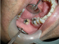
    Figure 1:  Intraoral view of missing 47 and abutment restored with silver
amalgam.
The second patient was a young, 25 years old, who wants a conservative replacement of missing posterior tooth as well as much concerned about the esthetics also (Figure 2). So a metal free Zirconia FDP was planned (Figure 3).
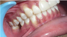
Figure 2: Intraoral Occlusal view showing missing 46.
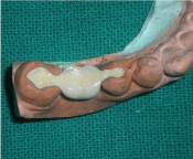
Figure 3: Zirconium FDP on master cast.
Clinical procedure
After the careful removal of the old restoration, the preparations were modified with proximal extension in the form of proximal box, with tapered round bur. All the line angles of occlusal and proximal preparation should be round with occlusally divergent walls. The finish line of the proximal box should be supragingival to minimize the chances of periodontal inflammation.
The final impressions were made by two step putty wash technique by using light body and putty consistency polyvinyl siloxane (Aquasil soft putty, Dentsply, Germany) (Figure 4). The impressions were poured in type IV dental stone (Ultrarock, Kalabhai Karson Pvt.ltd. Mumbai, India) to obtain the master cast (Figure 5).
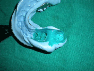
Figure 4: Final impression made by two step putty wash technique.
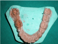
Figure 5: Master cast-Occlusal and proximal preparation.
After bite registration (Ramitec-Polyether bite registration material, 3M EPSE), the inlay cavities were filled with zinc oxide non eugenol cement. The wax pattern for all metal inlays FDP was fabricated with inlay wax and casted in all metal alloys. The casting was finished polished and fitting was properly evaluated before cementation.
The zirconia framework was fabricated by CAD/CAM procedure (Procera, Nobel Biocare, swizerland). The framework was properly evaluated for intraoral fitting and shade matching.
After proper evaluation of complete seating and marginal adaptation of the retainers, tissue relationship of the sanitary pontics and retainers of the both framework, the required Occlusal adjustments was done both in maximum intercuspation as well as in eccentric position to remove any premature contact with a fine diamond and carbide bur.
The permanent cementation of all metal inlays FDP was done with luting glass ionomer cement (Hy-bond Glasionomer CX, Shofu INC, Japan) after proper isolation (Figure 6). A layer of bonding agent (Prime and Bond NT, Dentsply, Germany) was applied over the margin to form a barrier to prevent the desiccation and dissolution of glass ionomer cement at the margin. After initial set, the extruded GIC was removed with curette. Figure 7 shows the all metal inlay FDP in occlusion.
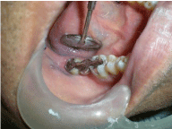
Figure 6: Intra oral view after cementation of inlay FDP.
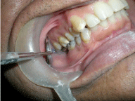
Figure 7: Inlay FDP in occlusion.
For cementation of Zirconia FDP, the etching of the enamel was done with 37% phosphoric acid for 20 seconds. After through irrigation with water spray and drying, the bonding agent was applied with a microbrush, uniformly spread with gentle air spray before curing for 20 seconds. To improve the mechanical interlocking, the fitting surface of the zirconia inlay retainers were sandblasted with 50 um alumina particles, instead of etching with 10% hydrofoluric acid which is not very effective.
The dual cure resin cement (Allcem, FGM, Brazil) was dispensed directly into the inlay cavities as well onto the fitting surface of the inlay retainers. The inlay FDP was placed gently with figure pressure, hold it in place after complete seating (Figure 8). All the extruded cement was removed before the polymerization. The light cure polymerization was done for 60 seconds with quartz tungsten halogen light curing unit. The resin cement in deeper part set by chemical polymerization. The margins were finished and polished with diamonds burs, rubber points and diamond polishing paste. Figure 9 shows the Zirconia inlay FDP in occlusion. During 5 and 3 year follow up of all metal and zirconia FDP respectively, both patients were satisfied with the outcome and no debonding was reported.

Figure 8: Intraoral view after cementation of zirconia FDP.

Figure 9: Inlay FDP in occlusion with sanitary pontic.
Discussion
Resin bonded FDP, Fiber reinforced composite resin FDP and inlay FDP are the conservative prosthesis considered for the replacement of the missing teeth under selected clinical conditions [5,6]. The resin bonded and fiber reinforced FDP’s can be successfully used as an alternatives for replacement of missing anterior teeth in young patients when conventional FPDs are contraindicated or when patient desires a conservative prosthesis. The conservative preparation, advancement in bonding systems and reported success suggest that this prosthesis can be used as a long term definitive alternative in specific clinical conditions for the replacement of missing anterior teeth but high incidence of debonding in posterior region preclude their use for the replacement of posterior tooth.
The all metal, Zirconia and PFM inlay FDP can be successfully considered as a minimum invasive technique for the replacement of single missing posterior tooth. Long term success depends on proper abutment selection, occlusal and proximal preparation, pontic design, careful bonding technique and type of occlusion.
Although, the zirconia and PFM prostheses are esthetically more superior than all metal FDP but ceramic causes the wearing of the enamel of opposing teeth as well as fracture of ceramic might occur which is absent in all metal FDP’s.
The extension of the occlusal preparation on to the proximal surface of the abutments in the form of box is essential to improve the strength and durability of the connectors as well as increase the mechanical resistance to any lingual, buccal and gingival displacement. The supragingival margins of the proximal box improve the periodontal health.
The sanitary pontic design is ideal design for replacing mandibular posterior tooth which improve the prognosis as patient is able to maintain good oral hygiene.
This technique is simple, easy and less time consuming than other approaches. It is an affordable and quick solution for the patients who reject more invasive treatments.
Summary
Under selected clinical conditions, inlay FDP can be considered as a conservative minimum invasive treatment modality for the replacement of single missing posterior tooth as an alternative to implant supported crown, conventional FDP and resin bonded FDP. The long term longitivity of the prosthesis depends on careful case selection, tooth preparation, material used for the fabrication of the inlay FDP and proper occlusal adjustments.
References
- Krejci I, Boretti R, Giezendanner P, Lutz F. Adhesive crowns and fixed partial dentures fabricated of ceromer/FRC: clinical and laboratory procedures. Pract Periodontics Aesthet Dent. 1998; 10: 487-498.
- Freilich MA, Duncan JP, Meiers JC, Goldberg AJ. Preimpregnated, fiberreinforced prostheses. Part I. Basic rationale and complete-coverage and intracoronal fixed partial denture designs. Quintessence Int. 1998; 29: 689- 696.
- Berekally TL, Smales RJ. A retrospective clinical evaluation of resin-bonded bridges inserted at the Adelaide Dental Hospital. Aust Dent J. 1993; 38: 85- 96.
- Hansson O, Bergstrom B. A longitudinal study of resin-bonded prostheses. J Prosthet Dent. 1996; 76: 132-139.
- Aboush Y, Estetah N. A prospective clinical study of a multipurpose adhesive used for the cementation of resin bonded bridges. Oper Dent. 2001; 26: 540- 545.
- Edelhoff D, Spiekermann H, Yildirim M. Metal-free inlay-retained fixed partial dentures. Quintessence Int. 2001; 32: 269-281.