Case Report
Austin J Clin Case Rep. 2016; 3(6): 1110.
A Case of Emphysematous Cholecystitis in a Patient with Diabetes Mellitus and Advanced Ovarian Cancer
Zaki W, Abujkeim N, Alawad AM*, Ramadan A, Abudames A, Ibrahim R and Tawfik S
Department of Surgery, Prince Sultan Armed Forces Hospital, Saudi Arabia
*Corresponding author: Awad Ali M. Alawad, Department of Surgery, Prince Sultan Armed Forces Hospital, Medina, Saudi Arabia
Received: November 28, 2016; Accepted: December 28, 2016; Published: December 30, 2016
Abstract
Acute emphysematous cholecystitis is a relatively rare disease, a severe variant of acute cholecystitis that predominantly affects elderly diabetic patients. Prompt diagnosis of emphysematous cholecystitis is critical, and the standard treatment is emergent cholecystectomy. In severely ill patients, percutaneous cholecystostomy with broad-spectrum antibiotics may be an alternative choice for treatment. We describe a diabetic woman with advanced ovarian cancer who developed septic shock due to acute emphysematous cholecystitis with rapid deterioration within 6 hours led to death.
Keywords: Emphysematous cholecystitis; Percutaneous cholecystostomy; Septic shock
Introduction
Emphysematous Cholecystitis (E.C) is an uncommon variant of acute cholecystitis in which the causative organisms are gas-forming bacteria. E.C has been defined clinically by the imaging demonstration of air in the gallbladder lumen; in the wall, or in the tissues adjacent to the wall of the gallbladder; and elsewhere in the biliary ducts in the absence of an abnormal communication with the gastrointestinal tract [1]. E.C is pathophysiologically different from acute or chronic cholecystitis. Obstruction of the gallbladder neck secondary to cholelithiasis induces acute and chronic cholecystitis. However, E.C mostly results from thrombosis or occlusion of the cystic artery with ischemic necrosis of the gallbladder wall. Diabetes mellitus is also a confounding factor for emphysematous cholecystitis [2].
The mortality rate from emphysematous cholecystitis is around 15%, higher than that of typical uncomplicated cholecystitis (1.4%) [3]. Due to the high mortality rate, prompt diagnosis and intervention are imperative. Plain radiography, ultrasonography, and computed tomography of the abdomen can provide much information for early diagnosis of this disease. The aim of this article is to present a case of EC and to attempt to elucidate the clinical entity and management of emphysematous cholecystitis.
Case Presentation
A 61-year-old woman with a history of diabetes mellitus, hypertension, and advanced ovarian cancer presented to the emergency department with right upper quadrant pain and vomiting for 4 days. She was diagnosed as a case of advanced ovarian cancer1 year ago, underwent bilateral oophorectomy and received adjuvant chemotherapy. The tumor recurs 1 month ago and she was scheduled for another chemotherapy regimen. On physical examination, she looked ill, her blood pressure was 160/85 mmHg, pulse rate 104 beats/minute, body temperature 39.2°C. Lung and heart sounds were normal. The abdomen was soft to palpation with bowel sounds present; however, there was mild upper-right quadrant tenderness without guarding, detectable hepato-splenomegaly, or ascites. Auscultation of the abdomen showed normal peristalsis. On admission, blood tests revealed total bilirubin level of 9.9 μmol/L (direct 6.3 μmol/L); Na 132 mmol/l and K 3.8 mmol/l. The white blood count was 29400/ μl with 94.8% neutrophils. Both abdominal radiograph and bedside ultrasonography showed no significant finding apart from the presence of gallstones.
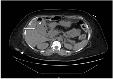
Provisional diagnosis of acute cholecystitis was made. The patient was admitted to ICU for close monitoring. The patient was treated empirically with ciprofloxacin and metronidazole, and intravenous administration of crystalloids. During the subsequent two hours following admission, the patient developed unexplained sudden cardiac arrest. She was resuscitated, sedated and intubated for ventilatory support. CT scan abdomen was done after stabilization and showed characteristic gallbladder distention, a circumferential gallbladder wall gas lucency, and an intraluminal air (Figure 1). Acute emphysematous cholecystitis was highly suspected then.

    Figure 1:  Abdominal computed tomography showing an enlarged gall bladder
with intraluminal (indicated by arrowhead) and intramural air (indicated by
white arrow).
Prompt cholecystectomy was considered to imply a very high risk under the circumstances of poor cardiac status. The patient underwent ultrasound-guided percutaneous cholecystotomy. Bile for culture revealed E. coli. The patient continued to deteriorate clinically despite the multiple therapeutic modalities in place. Unfortunately, she died on the fifth day after admission.
Discussion
To our knowledge, this is the first report that shows emphysematous cholecystitis in a diabetic patient with advanced ovarian cancer. Acute emphysematous cholecystitis is a relatively rare disease, a severe variant of acute cholecystitis, which exists with several differences compared with simple acute cholecystitis. It predominantly affects elderly men (men forming 71% of patients with emphysematous cholecystitis but only 27% in acute cholecystitis) [4]. A high incidence of diabetes mellitus (up to 50%) and high frequency of acalculous cholecystitis were noted too.
Radiological evaluation including plain abdominal radiograph, abdominal ultrasonography, and computed tomography of abdomen is the cornerstone of diagnosis of acute emphysematous cholecystitis [5]. Because ultrasonography of the gallbladder is the mainstay of diagnosis of gallbladder pathology, it is important to understand its limitations in the detection of emphysematous cholecystitis. This makes the early diagnosis of acute emphysematous cholecystitis challenging for any emergency physician. In our case, the initial abdominal radiograph and ultrasound were unremarkable apart from gallstones. CT is the best technique for diagnosing EC because it shows the exact location of air, whether in the gallbladder wall, in the gallbladder lumen, or throughout the bile duct, but it is irrational to perform CT for all patients with vague abdominal symptoms. However, high clinical suspicion with a radiological workup is important to reach the diagnosis.
Urgent cholecystectomy is the standard treatment for emphysematous cholecystitis. As an alternative technique percutaneous cholecystotomy may be used, if the patient’s situation is not suitable for surgical treatment [6]. The mortality rate for uncomplicated acute cholecystitis is approximately 1.4%. The mortality rate for acute emphysematous cholecystitis, however, is 15% to 20%, owing to the increased incidence of gallbladder wall gangrene and perforation in these patients [7]. In our case, there was sudden rapid deterioration due to septic shock that eventually ended with death despite early intervention. The presence of advanced malignancy and diabetes mellitus probably aggravated the condition.
Conclusion
The mortality rate for acute emphysematous cholecystitis is high. This variance emphasizes the importance of prompt diagnosis and emergent surgical intervention. Careful clinical monitoring and early surgical intervention may be the keys to reducing mortality.
References
- Chiu HH, Chen CM, Mo LR. Emphysematous cholecystitis. Am J Surg. 2004; 188: 325-326.
- Elsayes KM, Menias CO, Sierra L, Dillman JR, Platt JF. Gastrointestinal manifestations of diabetes mellitus: spectrum of imaging findings. J Comput Assist Tomogr. 2009; 33: 86-89.
- Gonzalez Valverde FM, Gomez Ramos MJ, Vazquez Rojas JL. Emphysematous cholecystitis. Clin Gastroenterol Hepatol. 2007; 5: e9.
- Khare S, Pujahari AK. A rare case of emphysematous cholecystitis. J Clin Diagn Res. 2015; 9: PD13-14.
- Narese F, Virzi V, Narese D, Sciortino AS, Orlando A, Culmone G, et al. Emphysematous cholecystitis: Imaging findings. Clin Ter. 2013; 164: e519- 522.
- Papavramidis TS, Michalopoulos A, Papadopoulos VN, Paramythiotis D, Karadimou V, Kokkinakis H, et al. Emphysematous cholecystitis: a case report. Cases J. 2008; 1: 73.
- Safioleas M, Stamatakos MK, Mouzopoulos GJ, Tziortzis G, Chagiconstantinu K, Revenas K. Emphysematous cholecystitis. Review of five cases and report of septic musculoskeletal complications. Chirurgia (Bucur). 2006; 101: 61-64.