Case Report
Austin J Clin Case Rep. 2017; 4(3): 1124.
A Case of Concomitant Peripheral Arterial Obstructive Disease and Polymyalgia Rheumatica
Girardi L*
Gesundheitszentrum Wien-Mariahilf, Austria
*Corresponding author: Girardi L, Gesundheitszentrum Wien-Mariahilf, Mariahilferstraße 85-87, A-1060 Vienna, Austria
Received: July 05, 2017; Accepted: August 17, 2017; Published: September 08, 2017
Abstract
A case of a patient with peripheral arterial obstructions is presented. We identified two conditions associated with vascular pathologies: diabetes mellitus and polymyalgia rheumatica. The findings, the course of the disease, and the response to cortison therapy taken into consideration, we reckon that both of these underlying conditions concomitantly caused the arterial changes: Proximal (femoral and popliteal) obstructions resolved with cortisol therapy, an angioplasty was performed for two infragenual lesions.
Keywords: Peripheral arterial disease (PAD); Diabetes mellitus; Atherosclerosis; Polymyalgia rheumatic (PMR); Giant cell arteritis (GCA)
Abbreviations
PAD: Peripheral Arterial Disease; GCA: Giant Cell Arteriitis; PMR: Polymyalgia Rheumatica; MS: Metabolic Syndrome; DM: Diabetes Mellitus
Introduction
The peripheral arterial disease (PAD) is in most cases caused by atherosclerosis which itself is in a large number of cases caused by diabetes mellitus (DM), i.e. the metabolic syndrome (MS). However, the giant cell arteritis (GCA), closely associated with the polymyalgia rheumatica (PMR) can also cause arterial obstructions. Although PAD and GCA are two distinct pathological entities, there is an increased risk for atherosclerosis in GCA patients.
Case Presentation
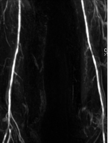
The 62 year old female patient presented with bilateral calf claudication after a walking distance of 100 to 200 meters over the last six months. It began on the right side, and was still felt more intensely and was lifestyle limiting on the right leg. No typical ischemic resting pain was reported, and there was no ulceration. The patient also complained of generalized muscle pain which she associated with the intake of a statin. Night sweats were reported, but no weight loss or elevated body temperature. The disease history included hypertension, hyperlipidaemia, osteoporosis, which were all medically treated, and a venous surgery. She had been smoking cigarettes until sometime earlier. The ankle-brachial-index was initially reduced on the right and within normal range on the left side. The Colour Doppler sonography and the Magnetic Resonance Imaging revealed moderate proximal bilateral stenosis in the superficial femoral arteries (more pronounced on the left side) and the left popliteal artery as well as relevant stenosis in the right tibiofibular trunk and in the right proximal posterior tibial artery. The peripheral arterial obstructive disease was diagnosed. Furthermore, carotid plaques were detected by sonography with a 50% stenosis in the left internal carotid artery. Aspirin (100 mg o.d.) was started in addition to the existing medication.
In the subsequent laboratory exams we diagnosed type 2 diabetes mellitus. The elevated sedimentation rate (30 mm/1h) in combination with the muscle pain led to the diagnosis of PMR, respectively GCA when vascular changes taken into consideration. A vessel biopsy was not performed due to the typical initial presentation combined with an elevated sedimentation rate and a good subsequent response to the corticosteroid therapy. The PMR was treated with tapering corticosteroid doses for about a year and a half, for DM the patient received metformin. The statin was replaced by ezetimibe (Figures1,2 and 3).

Figure 1: Magnetic resonance imaging at the time of diagnosis.

    Figure 2:  Digital subtraction angiography of the left superficial femoral artery
following corticosteroid treatment.

    Figure 3:  Digital subtraction angiography of the right superficial femoral
artery following corticosteroid treatment.
The patient was willing to undergo an angioplasty because the claudication was limiting her daily routine, but we waited for the PMR symptoms to resolve. At the beginning we attributed all vascular lesions in the legs to diabetes because the patient seemed to differentiate between the claudication and the generalized muscle pain and the claudication had had an earlier onset. Six months after the diagnosis the claudication was less severe, but still limiting on the right leg. At this time the ankle-brachial index was reduced on both legs. The decision was reached to perform an arterial angiography with possible angioplasty on both legs, delaying it yet for further tapering of the corticosteroid dose. After one and a half years following the initial presentation in our clinic a peripheral balloon angioplasty was performed on the right posterior tibial artery and the tibio-fibular trunk. Angiogram of the right and left femoral and popliteal arteries showed no relevant stenosis any more. The sonography performed after the angioplasty showed likewise no relevant stenosis in the right and left femoral and popliteal arteries, but a haemodynamically not relevant residual stenosis in the right tibio-fibular trunk after the balloon angioplasty. The ankle-brachial index, which was initially reduced on both legs, was now bilaterally normal. The patient became asymptomatic in regard to the PAD after the angioplasty and by this time had no more symptoms of the PMR.
Discussion
We present a case of peripheral arterial disease which can be attributed to two causes: atherosclerosis associated with diabetes mellitus and the giant cell arteritis associated with polymyalgia rheumatica. Patients with PMR, which is closely associated and overlapping with the GCA, have an increased risk of developing atherosclerotic PAD [1,2]. It has been postulated that the correlation is due to premature atherosclerosis caused by chronic inflammation, a known risk factor for atherosclerosis, or alternatively the arterial obstructions are manifestations of (subclinical) vasculitis [1]. Interestingly, a more recent study found no increased risk of coronary artery disease in patients with GCA [3]. A correlation between DM and GCA is not so clear: one study found a lower risk of DM in GCA patients [4], in another study risk factors were less atherogenic at incidence of GCA [3], yet another study found that DM increased the risk of GCA [5].
However, the vasculitic changes on large vessels in PMR can be a cause of arterial obstructions [6,7]. In our patient, retrospectively, the stenoses in the femoral and popliteal arteries were probably caused (mainly) by the GCA, the subgenual obstructions probably by diabetes. The claudication on the left leg might have been a part of the generalized pain caused by PMR (especially because the ankle-brachial index was normal at the first presentation), but it was described as a typical vascular claudication and resolved at the time when the anklebrachial index returned to normal. As the claudication on the right leg resolved promptly after the angioplasty was performed, it seems safe to assume that it was caused mainly by the obstruction of the tibiofibular trunk. We delayed the intervention because of the unclear association of the vascular changes to atherosclerosis and GCA, and indeed the proximal lesions resolved on corticosteroids alone. In case of subclavian arteries it has been described that an intervention can be avoided by waiting for the results of the corticosteroid therapy [8].
At the initial presentation the patient was on statin therapy which was substituted by ezetimib because statins can cause muscular pain. Statin exposure was not associated with the GCA, yet could favour the tapering of the corticosteroid doses [9]. Nevertheless we did not change this medication later on.
References
- Warrington KJ, Jarpa EP, Crowson CS, Cooper LT, Hunder GG, Matteson EL, et al. Increased risk of peripheral arterial disease in polymyalgia rheumatica: a population-based cohort study. Arthritis Res Ther. 2009; 11: R50.
- Nwadibia U, Larson E, Fanciullo J. Polymyalgia Rheumatica and Giant Cell Arteritis: A Review Article. S D Med. 2016; 69: 121-123.
- Udayakumar PD, Chandran AK, Crowson CS, Warrington KJ, Matteson EL. Cardiovascular risk and acute coronary syndrome in giant cell arteritis: a population-based retrospective cohort study. Arthritis Care Res (Hoboken). 2015; 67: 396-402.
- Ungprasert P, Upala S, Sanguankeo A, Warrington KJ. Patients with giant cell arteritis have a lower prevalence of diabetes mellitus: A systematic review and meta-analysis. Mod Rheumatol. 2016; 26: 410-414.
- Abel AS, Yashkin AP, Sloan FA, Lee MS. Effect of diabetes mellitus on giant cell arteritis. J Neuroophthalmol. 2015; 35: 134-138.
- Buttgereit F, Dejaco C, Matteson EL, Dasgupta B. Polymyalgia Rheumatica and Giant Cell Arteritis: A Systematic Review.JAMA. 2016; 315: 2442-2458.
- Narváez J, Estrada P, López-Vives L, Ricse M, Zacarías A, Heredia S, et al. Prevalence of ischemic complications in patients with giant cell arteritis presenting with apparently isolated polymyalgia rheumatica. Semin Arthritis Rheum. 2015; 45: 328-333.
- Van der Matten B, Zandbergen AA, Dees A. Bilateral axillary arterial obstruction. BMJ Case Rep. 2014.
- Pugnet G, Sailler L, Bourrel R, Montastruc JL, Lapeyre-Mestre M. Is statin exposure associated with occurrence or better outcome in giant cell arteritis? Results from a French population-based study. J Rheumatol. 2015; 42: 316- 322.