Surgical Case Report
Austin J Clin Case Rep. 2018; 5(1): 1127.
Placing an Orientation Suture during an Elliptical Excision Procedure is Simple, Efficient, Safe, and may Help Preserve Tissue Integrity
Hnath GJ¹, Xia Y²* and Cho S³
¹Naval Hospital, Portsmouth University, USA
²Dermatology Service, San Antonio Military Medical Center, USA
³Dermatology Service, Tripler Army Medical Center, USA
*Corresponding author: Xia Y, Dermatology Service, San Antonio Military Medical Center, 3551 Roger Brooke Dr. San Antonio, USA
Received: January 09, 2018; Accepted: February 02, 2018; Published: February 14, 2018
Abstract
The views expressed in this article are those of the authors and not necessarily reflect the official policy or position of the Department of the Navy, Department of the Army, Department of Defense, or the United States Government.
An elliptical skin excision is often performed to provide diagnostic information on managing cutaneous disorders. We propose a simple and safe modification of the standard technique by placing an orientation suture in the specimen before it is removed from the skin in order to provide traction and prevent structural damage to the tissue.
Case Presentation
An elliptical skin excision is often performed as part of an incisional or excisional biopsy to provide diagnostic information for managing cutaneous disorders [1]. In addition to establishing or confirming a diagnosis, an elliptical skin excision can be used to evaluate the effectiveness of therapy, provide a specimen for histological assessment of margins, and offer a surgical cure for numerous cutaneous disorders [2].
Commonly, an elliptical excision is performed after the surgical site has been marked, anesthetized, prepped, and draped [3]. In the standard procedure, lateral margins of the excision are made, the elliptical specimen is grasped at one of its poles with a pair of tissue forceps and the tissue is elevated. The surgeon then separates the specimen from underlying tissues along a horizontal, even plane using a scalpel or sharp tissue scissors [2]. A suture to remind the surgeon of the orientation is often placed after the specimen has been completely removed from the underlying tissue. The specimen is then transferred to the appropriate fixative container and subsequently submitted to pathology.
We propose a simple and safe modification of the standard technique by placing the orientation suture in the specimen before it is removed from the skin in order to provide traction during the removal process without compounding structural damage to the tissue.
Technique
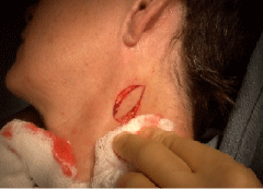
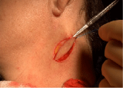
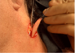
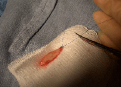
After incising the lateral borders of the intended specimen, an orientation suture is placed at a defined pole (usual the distal end) of the ellipse while the specimen is still in situ. We recommend using either dissolvable or non-dissolvable sutures that are already available for closure of the wound. The suture ends are cut long (3 cm or greater) and grasped with the fingers of the non-dominant hand. Gentle traction is then applied using the suture ends to elevate the tissue (Figure 1- 4). This facilitates further dissection with a scalpel or tissue scissors to free the specimen from the underlying tissues in the usual fashion. The specimen is then easily transferred into the specimen container.

Figure 1: The border of the specimen is freed from the lateral skin.

Figure 2: An orientation suture is placed at superior pole of the specimen.

    Figure 3:  Gentle traction is placed on the orientation suture as the specimen
is excised.

    Figure 4:  The resulting specimen is completed excised with the orientation
suture placed at the superior pole.
Discussion
This modification of a standard biopsy technique offers several advantages. By using a marking suture as a surgical aid in this manner, the surgeon simultaneously provides a means for specimen dissection, while helping to ensure proper orientation. This technique also helps maintain the integrity and architecture of the specimen by decreasing crush or tear artifacts often caused inadvertently by forceful use of forceps. It also improves time and procedural efficiency by decreasing the steps of specimen excision and the number of surgical instruments handled. There should be no additional cost to using the described technique as sutures already available for the closure will be utilized.
In short, this technique provides a quick, easy, cost-effective, and safe method for dissecting excisional specimens while reducing artifactual tissue injury and orientation errors.
References
- Farmer ER. Why a skin biopsy? Arch Dermatol. 2000; 136: 779-780.
- Garcia C. Skin biopsy techniques. In: Robinson JK et al: Surgery of the skin. Philadelphia: Elsevier Mosby; 2005.
- Robinson JK. Elliptical incisions and closures. In: Robinson JK et al: Atlas of cutaneous surgery. Philadelphia: WB Saunders; 1996.