Clinical Image
Austin J Clin Case Rep. 2018; 5(1): 1127.
End Stage of Ogilvie Syndrome
Valentim M*, Ramalho J, Almeida S and Gameiro A
Department of Internal Medicine, Hospital Distrital de Santarém, Portugal
*Corresponding author: Valentim M, Avenida Bernardo Santareno, Santarém, Portugal
Received: December 08, 2017; Accepted: February 02, 2018; Published: February 14, 2018
Clinical Image
A 87-year-old female, with several episodes of bowel pseudoobstruction in the last 4 years, with no apparent cause; was admitted to the emergency department for abdominal pain and distension for the last 2 days.
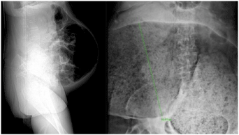
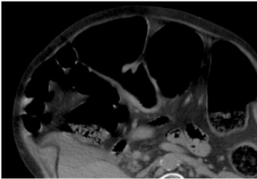
On presentation, temperature was 38.4°C and blood pressure 93/40 mmHg. The abdominal examination revealed: murmur abolished, volumosos distension and tympanic sound over all portions. Laboratory finding showed: hemoglobin 11.2 g/dL, leukocytosis 41.5 x 10⁹/L with 96.7% neutrophilia and C-reactive protein 27.79 mg/dL; with the following abdominal X-Ray (Figure 1) and CT-scan (Figure 2).
In the first 24 hours a conservative management was decided: correct fluids and electrolytes, Nil per os, nasogastric and rectal tube suction, IV metoclopramida and IV Neostigmine, and after stabilization perform a Colonoscopic decompression of the colon.
Progressive deterioration of the clinical condition with multiple organ failure, dying 24 hours after admission.

