Case Report
Austin J Clin Case Rep. 2019; 6(2): 1145.
Giant Splenic Pseudocyst: A Case Report and Literature Review
Feng Y, Qi X, Jin Y, Yu Y, Geng H and Li J*
Department of General Surgery, The Second Affiliated Hospital, School of Medicine, Zhejiang University, China
*Corresponding author: Jiangtao Li, Department of General Surgery, The Second Affiliated Hospital, School of Medicine, Zhejiang University, 88 Jiefang Road, Hangzhou City, Zhejiang Province, China
Received: April 23, 2019; Accepted: May 27, 2019; Published: June 03, 2019
Abstract
Splenic cyst is a rare clinical disease, which is commonly found for young people. It is asymptomatic in the early stage, and often identified by physical examination after with compression symptoms due to cyst enlargement presented at the later stage. The treatment is varied due to extremely low incidence rate. A case of a patient with a giant splenic pseudocyst was presented, the experience of diagnosis and treatment for this case and literatures was summerized.
Keywords: Splenic cysts; Pseudocysts; Splenectomy
Introduction
Splenic cysts are an extremely rare clinical condition that occurs in about 0.07% of the population [1]. According to Marin, splenic cysts can be classified into true cysts or pseudocysts based on the presence or absence of epithelial lining in the cyst wall [2]. Splenic pseudocysts are often formed by post-injury spleen pulmonary hematoma or limited liquefaction following splenic infarction [3], and are commonly found below the splenic capsule. Patients with splenic cysts generally exhibit no specific clinical symptoms, and these cysts are often found accidentally during imaging examination. Ultrasound and computed tomography (CT) can be used for the preliminary clinical diagnosis, but definitely diagnose cannot be concluded with either way. The main treatment including aspiration, cyst fenestration and drainage, partial splenectomy and total splenectomy. However, it is difficult to choose the suitable treatment due to limited evidence. we presented a case of giant splenic cyst and summerized the results of previous studies (Table 1) so as to get more understanding for this disease.
Author
Country
Year
Therapy
Vuyyuru S[6]
USA
2017
Uneventful hand-assisted aparoscopic
Kumar P[7]
India
2016
Laparoscopic splenectomy
Ozlem N[8]
Turkey
2015
Splenectomy
2014
Splenectomy
Rana SS[9]
India
2014
Endoscopic transpapillary drainage
Garg M[10]
India
2013
Splenectomy
Gilani SM[11]
USA
2013
Exploratory laparotomy and splenectomy
Forouzesh M[12]
Iran
2013
Exploratory laprotomy with splenectomy
Ingle SB[13]
India
2013
Emergency therapeutic splenectomy
Yildiz P[14]
Turkey
2013
Elective surgery
Ozkan F[15]
Turkey
2013
Laparoscopic partial cystectomy and Omentoplasty but preserve the spleen
Pukar MM[16]
India
2013
Radical en bloc splenic resection (together with resection of the diaphragm and subcutaneous tissue).
Table 1: The case report statistics about cyst of spleen.
Case Presentation
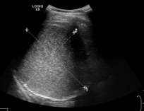
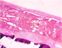
A 42-year-old male patient was admitted to our hospital upon “spleen-occupying mass by physical examination for one week”. He was healthy and had no history of cardiovascular disease and trauma. He had no positive sign for physical examination. Laboratory test was in the normal range. Ultrasound revealed a giant cystic mass in the spleen, about the size 11 x 13 x 12 cm, that was initially diagnosed to be an epidermoid cyst (Figure 1). Enhanced CT scan showed a giant splenic cyst with calcification of cyst capsule (Figure 2). The patient underwent laparoscopic splenectomy. A smooth and intact capsule was visible around the spleen during the operation. A cystic mass approximately 12*13cm in size with partial calcification on the surface was identified within the spleen, after complete splenectomy, cystic fluid was aspirated for bacterial culture, and no general bacteria and fungi were identified. Postoperative histopathological examination indicated that the mass was a calcified splenic pseudocyst (Figure 3). The patient was uneventfully and discharged on postoperative day 5, and the results of abdominal ultrasound was normal postoperative on one month follow up.

    Figure 1: Ultrasound examination of the abdomen revealed a giant cystic
mass in the spleen, which was initially dignosed to be an epidermoid cyst.

    Figure 2: Enhanced CT imaging of the abdomen showed a giant splenic cyst
with calcification at the edges.

    Figure 3: Pathology results indicated that the mass was a calcified splenic
pseudocyst.
Discussion
Splenic cysts refer to the cystic lesions of the spleen. Due to the lack of specific clinical symptoms in the early stage, the vast majority of patients was found accidently. Various clinical imaging techniques such as ultrasound, CT and MRI is help to the preliminary diagnosis. Ultrasound examination can help determine of the nature of the cyst content, presence or absence of calcification in the cyst wall, presence or absence of septum within the cyst, and whether or not the cyst wall is regular. Magnetic Resonance Imaging (MRI) T2 images can display cystic fluid as a region of water-like high signal intensity. In addition, CT examination can further confirm the nature of the cystic fluid, morphology of the cyst, and the relationship between the cyst and surrounding tissues. However, neither of the above techniques can distinguish between true cysts and pseudocysts. Therefore, the gold standard for the clinical diagnosis of splenic cysts is histopathology. Although in pathology, splenic cyst is a benign disease, however, in some patients, the elevation of tumor markers in blood or cystic fluid, such as CA199 CA125 and CEA, but the underlying mechanism is unclear. Takamitsu Inokuma [4] hypothesized that these markers may be secreted in the cystic fluid by the epithelial cells, and upon cyst rupture, absorption of the cystic fluid by the peritoneum leads to an increase in the serum levels of the markers. This case had normal serum tumor marker and cystic fluid CEA.
Treatment for splenic cysts mainly includes splenic cyst aspiration, cyst fenestration and drainage, partial splenectomy and total splenectomy. Treatment regimens for splenic cysts are mainly selected based on patient’s age, size and location of cyst, presence or absence of cyst-induced compression symptoms, and presence or absence of cyst rupture, hemorrhage and infection. Generally, cyst aspiration and fenestration/drainage have poor outcome and can easily lead to cyst recurrence. Rana AP [5] proposed several treatments for symptomatic patients or those suspected with malignant cysts. Partial splenectomy and conservative treatment (cyst aspiration) are recommended for patients with splenic cysts less than 5cm in diameter. On the other hand, total splenectomy (open or laparoscopic splenectomy) is recommended for patients with larger splenic cysts to prevent cyst rupture, hemorrhage, infection and recurrence. For larger splenic cyst, complete excision of the specimen is more difficult, we recommend the first puncture and aspiration of the cyst, followed by splenectomy.
Conclusion
The goal of splenic cyst treatment is to relieve symptoms, prevent concurrent symptoms and cyst recurrence. The patient’s age, size and location of the cysts, and presence or absence of symptoms, cyst rupture, hemorrhage and infection should be considerable so as to provide the optimal treatment for such patient.
References
- Robbins FG, Yellin AE, Lingua RW, Craig JR, Turrill FL, Mikkelsen WP. Splenic epidermoid cysts [J]. Ann Surg. 1978; 187: 231-235.
- Martin JW. Congenital splenic cysts [J]. Am J Surg. 1958; 96: 302-308.
- Kishanchand C, Naalla R, Kumar S, Mathew M. Non-traumatic pseudocyst of spleen presenting as chronic abdominal pain and vomiting [J]. BMJ Case Rep. 2014; 2014, bcr2014207133.
- Inokuma T, Minami S, Suga K, Kusano Y, Chiba K, Furukawa M. Spontaneously Ruptured Giant Splenic Cyst with Elevated Serum Levels of CA 19-9, CA 125 and Carcinoembryonic Antigen [J]. Case Rep Gastroenterol. 2010; 4: 191-197.
- Rana AP, Kaur M, Singh P, Malhotra S, Kuka AS. Splenic epidermoid cyst - a rare entity [J]. J Clin Diagn Res. 2014; 8: 175-176.
- Vuyyuru S, Kharbutli B. Epidermoid cyst of the spleen, a case report [J]. Int J Surg Case Rep. 2017; 35: 57-59.
- Kumar P, Hasan A, Kumar M, Singh V. Isolated hydatid cyst of spleen: A rare case with rare presentation [J]. Int J Surg Case Rep. 2016; 28: 279-281.
- Ozlem N. Traumatic rupture of a splenic cyst hydatid [J]. Int J Surg Case Rep. 2015; 7C: 112-114.
- Rana SS, Chaudhary V, Sharma V, Sharma R, Dutta U, Bhasin DK. Infected pancreatic pseudocyst of spleen successfully treated by combined endoscopic transpapillary stent placement and transmural aspiration [J]. Gastrointest Endosc. 2014; 79: 360-361.
- Garg M, Kataria SP, Sethi D, Mathur SK. Epidermoid cyst of spleen mimicking splenic lymphangioma [J]. Adv Biomed Res. 2013; 2: 49.
- Gilani SM, Tashjian R, Qu H. Histopathological, histogenetic, and immunohistochemical analysis of epidermoid cyst of spleen [J]. Acta Chir Belg. 2013; 113: 325-329.
- Forouzesh M, Ghanbarzadegan L, Rahimi M, Ghahramani L. Splenic Epidermoid Cyst during Pregnancy; Case Report and Review of the Literature [J]. Bull Emerg Trauma. 2013; 1: 179-181.
- Ingle SB, Hinge CR, Jatal SN. An interesting case of primary epithelial cyst of spleen [J]. Indian J Pathol Microbiol. 2013; 56: 181-182.
- Yildiz P, Pasaoglu E, Behzatoglu K, Huq Erdem G, Bozkurt ER. Multiloculated epidermoid cyst of spleen: a case report. [J]. Turk Patoloji Derg. 2013; 29: 238-240.
- Ozkan F, Yesilkaya Y, Peker O, Yuksel M. Anaphylaxis due to spontaneous rupture of primary isolated splenic hydatid cyst [J]. Int J Crit Illn Inj Sci. 2013; 3: 152-154.
- Pukar MM, Pukar SM. Giant solitary hydatid cyst of spleen-A case report [J]. Int J Surg Case Rep. 2013; 4: 435-437.