
Clinical Image
Cadena I, Valentim M, Grego M and Siopa L. Constrictive Pericarditis: Steel Heart. Austin J Clin Case Rep. 2019; 6(3): 1149.
Constrictive Pericarditis: Steel Heart
Cadena I*, Valentim M, Grego M and Siopa L
Department of Internal Medicine, Hospital Distrital de Santarém, Portugal
*Corresponding author: Cadena I, Avenida Bernardo Santareno, Santarém, Portugal
Received: June 10, 2019; Accepted: June 22, 2019; Published: June 29, 2019
Clinical Image
Male 78 years old with history of atrial fibrillation, was evaluated for progressively worsening of dyspnea and edema of the legs. On presentation, he had mild dyspnea, hypotension, reduced vesicular murmur in the left lung base and pitting edema. A Chest x-ray: left pleural effusion in the lower 2/3, an electrocardiogram showed lowvoltage and chronic atrial fibrillation. The suspection de effusion pleural of unknown etiology motivated to the performance of diagnostic thoracentesis compatible with transudate and TAC-thorax showed diffuse thickening of the pericardium without associated effusion. Echocardiogram showed: thickened pericardium, with no significant effusion interpreting pericarditis. A catheterization, revealing an increase in the pressures of the right heart chambers. Constrictive Pericarditis was diagnosed and marked pericardial thickening was observed during a pericardiotomy. An analysis of the histological piece was reported as fibrotic alterations compatible with Constrictive Pericarditis, confirming the diagnosis, but no cause was identified (Figure 1).
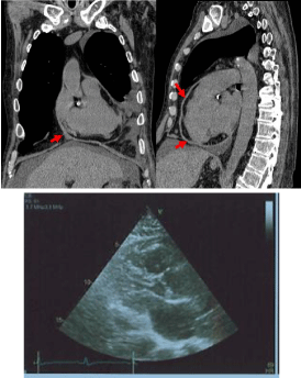
Figure 1: