
Clinical Image
Austin J Clin Case Rep. 2019; 6(5): 1160.
Post-ERCP Acute Necrotizing Pancreatitis
Cadena I*, Sara N, Fonseca S and Grego M
Department of Internal Medicine, Hospital Distrital de Santarém, Portugal
*Corresponding author: Cadena I, Avenida Bernardo Santareno, Santarém, Portugal
Received: December 19, 2019; Accepted: December 26, 2019; Published: December 31, 2019
Clinical Image
73-year-old woman was admitted to our hospital, due to clinical symptoms of abdominal pain, vomiting, jaudince and fever. Physical examination showed dehydrated, conjunctival jaundice and painful palpation in the right hypochondrium. The blood test showed: Leukocytes 14000x109/L Leukocytes, Total Bilirubin 2.0mg/dl, Direct Bilirubin 0.7mg/dl, AST 122U/L, ALT 150U/L. In the Abdominal Ultrasonography it was noted a slight hepatomegaly with a normal Gallbladder with no evidence of lithiasis or intrahepatic biliary ducts dilatation. Meanwhile in the common bile duct there was indirect signal of dilatation probable caused by Gallstones. An ERCP was performed and showed a dilated primary biliary tract with small and multiple small lacunar images, an extraction of gallstones were done. During admission 24 hours after-ERCP, the patient clinical condition worsened. The blood test evidenced leukocytosis of 22000 x109U/L, amylase 2087U/L, lipase 4624U/L and blood cultures grew an ESBLproducing E. Coli and Antibiotic therapy were performed. The CT abdomen revealed an increased dimensions of the head and body of the pancreas, with areas of edema and necrosis suggesting acute necrotizing pancreatitis (Panel A and B). Progressive deterioration of the clinical condition with multiple organ failure, dying 96 hours after admission.
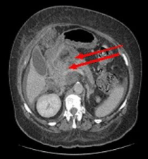
Panel A:
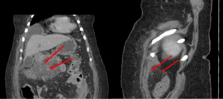
Panel B: