Case Report
Austin J Clin Case Rep. 2020; 7(3): 1173.
A Mysterious Intra-Cardiac Needle Discovered Accidentally During Pericardiothentesis under Fluoroscopy
Alkady H*, Abouramadan S and Labeeb D
Department of Cardiothoracic Surgery, Cairo University, Egypt
*Corresponding author: Hesham Alkady, Department of Cardiothoracic Surgery, Cairo University, Kasralaini str., Almanial, Cairo, Egypt
Received: May 22, 2020; Accepted: October 09, 2020; Published: October 16, 2020
Abstract
Due to the rarity of retained intra-cardiac sewing needles, no clear guidelines exist regarding the indication for their extraction. In this study we report a case of a sewing needle extracted from the right atrium of an adult female presenting with recurrent pericardial effusion after one year of accidental penetration.
Keywords: Intra-cardiac needles; Pericardiothentesis; Echocardiography; Cardiopulmonary bypass; Sternotomy
Introduction
Intra-cardiac sewing needles detected in adults and children may be due to accidental penetration e.g. while sleeping, self-inflicted as a result of mental and psychic disorders or domestic abuse [1,2]. Due to the rarity of retained intra-cardiac sewing needles, no clear guidelines exist regarding the indication for their extraction. Conservative or operative options should be individualized according to the timing of presentation (acute or delayed), presence of symptoms (e.g. chest pain, infection, arrhythmia) as well as location [3]. In this study we report a case of a sewing needle extracted from the right atrium of an adult female after one year of accidental penetration.
Case Presentation
A 60-year-old female patient was referred to our outpatient clinic due to a needle found by the cardiologist during pericardiothentesis under fluoroscopy for increasing pericardial effusion (Video).
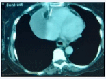
The clinical history, physical examination as well as laboratory studies of the patient were unremarkable apart from recent progressive shortness of breath since 2 months. Therefore, the cardiologist decided to do a pericardial aspiration with chemical, bacteriological and cytological examination of the pericardial fluid after increase its amount despite of diuretic therapy. The pericardial aspirate was sero-sanguinous exudative in nature, negative for organisms and malignant cells. The patient had no explanation how a needle reached this place and she did not receive any major procedure before could be the cause. The patient is also well-educated and her mental as well as psychological assessments were within normal. A transthoracic echocardiography showed an echogenic linear object at the wall of right atrium, unclear whether inside or outside the atrial cavity, as well as mild pericardial effusion with thickening of the pericardium. Multi-slice CT chest showed that the needle lies inside the right atrial cavity embedded in its wall (Figure 1).

    Figure 1: A Multi-slice CT of the chest showing the needle in the right atrial
cavity as well as collected pericardial effusion on its lateral surface. The
pericardium appears slightly thickened.
The possible hazards of leaving the needle in the heart were explained to the patient as well as the advantage of biopsying the pericardium during surgical extraction to determine the cause of recurrent effusion. The patient was first terrified from the surgical intervention; however, she consented surgery at the end.
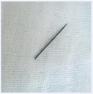
Through full median sternotomy, the thickened pericardium was opened and sero-sanguinous effusion was evacuated. Some adhesions were found on the anterior surface of the heart especially right atrium. Trials to locate the needle by digital palpitation of the right atrium were unsuccessful. A mobile C-arm X-ray machine was brought to the operation room to confirm the position of the needle inside the right atrial cavity. Cardiopulmonary was then initiated via aorto-bicaval cannulation. After snaring of both cavae, the right atrium was opened transversely along the atrio-ventricular groove and astonishingly a rusty sewing needle was found embedded with its sharp end in the roof of the right atrium near the tricuspid valve (Figure 2).

    Figure 2: The rusty sewing needle found in the cavity of the right atrium
embedded in its wall.
Upon seeing the sewing needle, the patient remembered that she brought that type of needles one year ago and lost one of them shortly after. So the only available explanation postulated is that the needle went unnoticed into the patient´s chest may be while sleeping nearby the needle or leaving it inside her pocket which then eroded its way through the pericardium onto the right atrium being an anterior structure immediately behind the chest wall. The postoperative recovery went uneventfully and the patient was discharged home safely after one weak. Pathological examination of the thickened pericardium revealed nonspecific chronic inflammation with no signs of malignancy.
Discussion
Upon reviewing the literature, there is a general tendency to remove intra-cardiac needles once diagnosed to avoid potential hazards or prevent further damage [4]. These hazards include embolization, thrombus formation, endocarditis, pericarditis and injury to cardiac structures including cardiac perforation and pericardial tamponade. [5] Hazards are more prone when the needles are located in left-sided chambers and when partially embedded in myocardium [6].
Localization modalities of intra-cardiac needles include echocardiography and more accurately computed tomography. Intraoperative C-Xray is much helpful and time sparing. Interventional extraction using modern radiological facilities would be difficult and may be hazardous. Median sternotomy is much superior than any other approaches e.g. thoracotomies as it allows the best exposure and safest access to cardiopulmonary bypass. Sometimes the needle can be removed without the help of the heart machine and sometimes not [7,8].
In our study, although the needle penetrated accidentally into the chest wall and then right atrium, yet it did cause some sort of chronic pericarditis and recurrent effusion one year later. This represented, beside the sharp nature of the non-sterile sewing needle, the indications to remove the needle.
Conclusion
Since the fact that no clear guidelines exist regarding the indication for extraction of intra-cardiac needles, therefore management whether conservative or operative should be individualized according to each case. Surgical removal of lately-discovered intra-cardiac needles should be considered if complications occurred like pericarditis causing recurrent pericardial effusion in our case.
References
- Talwar S, Subramaniam KG, Subramanian A, Kothari SS, Kumar AS. Sewing needle in the heart. Asian Cardiovasc Thorac Ann. 2006; 14: 63-65.
- Sola JE, Cateriano JH, Thompson WR, Neville HL. Pediatric penetrating cardiac injury from abuse: a case report. Pediatr Surg Int. 2008; 24: 495-497.
- Perrotta S, Perrotta A, Lentini S. In patients with cardiac injuries caused by sewing needles is the surgical approach the recommended treatment? Interactive CardioVascular and Thoracic Surgery. 2010; 10: 783-792.
- Ngaage DL, Cowen ME. Right ventricular needle embolus in an injecting drug user: the need for early removal. Emerg Med J. 2001; 18: 500-501.
- Inoue T, Iemura J, Saga T. Delayed cardiac tamponade caused by selfinserted needles. Can J Cardiol. 2003; 19: 306-308.
- Nishida S, Tomita S, Watanabe G, Yasuda T, Iino K, Arai S. Intramyocardial foreign body: sewing needle with the uncommon clinical feature of constrictive pericarditis. Jpn J Thorac Cardiovasc Surg. 2005; 53: 598-600.
- Sayin AG, Besirli K, Arslan C, Cantu¨rk E. A case of intramyocardial¸ sewing needle extracted without stopping the heart. Injury. 2002; 33: 276-277.
- Murakami M, Okada H, Nishida M, Hamano K. A sewing needle completely buried in the myocardium removed under extracorporeal circulation. Ann Thorac Cardiovasc Surg. 2006; 12: 216-218.