Case Report
Austin J Clin Case Rep. 2020; 7(3): 1175.
An Unusual Cause of Recurrent Abdominal Pain
Dev S, Yadav DP*, Shukla SK and Dixit VK
Department of Gastroenterology, Banaras Hindu University, India
*Corresponding author: Dawesh Prakash Yadav, Department of Gastroenterology, Sir Sundarlal Hospital, Institute of Medical Sciences, Banaras Hindu University, Varanasi – 221005, India
Received: May 24, 2020; Accepted: October 17, 2020; Published: October 24, 2020
Abstract
Lead poisoning has been recognized as a major public health risk. We present a case of middle aged female, who presented with recurrent episodes of severe abdominal pain, which remains obscure even after thorough clinical evaluation and extensive investigations. Lastly, blood lead levels came out to be significantly raised and diagnosis of lead intoxication was made convincingly. On reviewing the case, history of intake of herbal medication justified the diagnosis. Therefore, even though the diagnosis represents a challenge, a physician must always include this possibility in the differential diagnosis for cases with suggestive symptoms.
Keywords: Lead; Ras sindoor; Herbal medication; Pain abdomen
Introduction
Lead poisoning has been recognized as a major public health risk, particularly in developing Countries [1]. It may involve major organs. The following case report argument about GI presentation of lead toxicity.
Case Presentation
A 34 years old woman, Resident of North India and nurse by occupation presented to us with complaint of diffuse abdominal pain for last 3 days. Pain was acute onset, moderately severe in intensity, colicky, starting from lower abdomen, involving whole abdomen over few hours with increased severity. No precipitating and relieving factors were present. It was associated with non passage of flatus and stools for last 2 days, but was not associated with abdominal distention or vomiting.
Patient also gave history of similar episodes in last 2 months. No history of chronic drug intake, substance abuse or surgical intervention in past. She consulted a gynaecologist few months back for primary infertility, but no records were available.
On examination, vitals were stable and mild pallor was present per abdomen examination showed diffuse tenderness but no guarding or rigidity was present. Bowel sound were sluggish. Per rectal examination was normal.
Patient was admitted in Acute care unit and Urgent X ray Abdomen erect posture was performed which showed few dilated large bowel loops, but essentially ruled out perforation. She was started on conservative management in the form of restricted diet, IV fluids, enema and terpentine oil stooping.
Routine investigations showed moderate Anemia with Hemoglobin of 8 gm/dl (MCV- 72 fL) and low platelets (1.2 lakh/ mm3) with normal total leucocyte counts. Liver functions were also mildly deranged in form of transaminitis (SGOT/PT =72/67 IU/L) with normal renal functions and serum amylase/ lipase levels. CT Enterography was done which turned out to be normal.
Patient got improved with conservative measures after 2 days and was discharged on SOS pain killers and laxatives , to follow up for further evaluation.
Just 3 days after discharge , she again presented with similar nature of pain for last 2 days, not associated with vomiting / abdominal distention or non passage of flatus or stools. She was admitted and conservative management was started. Repeat CT Enterography and Angiography was done which was reported normal. Prepared full length Colonoscopy and Esophagogastroduodenoscopy were also performed on the following days, but were essentially normal. Routine investigations showed bicytopenia in form of Hb=9 gm/ dl and marginally low platelets. General blood picture showed predominantly microcytic cells with hypochromia and few cells showing basophilic stippling. Taking clue from that , serum lead levels was ordered , which came out to be raised (75μg/dl, five times the ULN). For making a consolidate diagnosis, it was repeated from another standard lab, which again came high (65μg/dl). Simultaneously urine porphyrin levels were done, which came out to be normal.
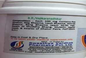
On reviewing the history , patient admitted to consumption of an ayurvedic preparation for last few months as an appetizer. On analyzing the preparation , it was found to contain Ras sindoor (Lead) as the principle ingredient . The suspected source of exposure in our patient was herbal-based medication (Figure 1). Though toxicological analysis was not performed, absence of any other source of poisoning and circumstantial evidence of herbal-based medicinal use, which have been widely reported to cause lead poisoning, supports our diagnosis.

Figure 1: Herbal Preparation containing Ras Sindoor (Lead).
We considered the toxicology references [2], and found that only oral chelator (DMSA/Succimer) was recommended for the patient. So, it was started at the recommended dose of 10mg/kg three times a day for five days, followed by 10 mg/kg twice a day for next two weeks (maximum 500 mg/dose) . Patient improved symptomatically and repeated serum lead levels were also reported within the normal range.
Discussion
Common sources of lead exposure include lead paint, lead– acid batteries, soil contamination near factories, lead soldering, cosmetics and herbal-based medications [3].
The ways of contamination include ingestion, inhalation, prenatal exposure, and dermal exposure, but the most important and frequent ones are ingestion and inhalation [4]. The half-life of lead is between 30 and 40 days in human body and it binds to the sulfhydryl group of proteins leading to toxicity for multiple enzyme systems [5].
The clinical presentation of lead poisoning involves nervous, hematologic, and renal systems impairment, but it can also lead to gastrointestinal disorders (anorexia, vomiting, constipation, colicky abdominal pain), hypertension, and fertility impairment [6]. Neurological symptoms include foot/wrist drop, ataxia, stupor, coma, convulsions, hyperirritability. Oral examination commonly reveals the Burtonian line on gum [7]. Impairment of the hematological system may involve either disruption of heme synthesis or hemolysis, leading to Basophilic stippling with microcytic hypochromic anaemia and thrombocytopenia has also been reported.
The effects of lead on the renal system consist of proximal tubular function impairment leading to aminoaciduria, glycosuria [8], and hyperphosphaturia, interstitial nephritis in chronic exposure, and also impairment of calcium metabolism by interfering with activation of vitamin D 1,2- dihydroxy cholecalciferol.
The findings pertaining to lead poisoning in our patient were - Recurrent abdominal colics, anaemia, thrombocytopenia and deranged transaminases.
The diagnosis is established on the basis of blood lead levels higher than 10μg/dl1. If a patient is found with high blood lead level, the test must be repeated before considering any therapy. Chelating agents are recommended only if the level is above 45μg/dL, and the type should be chosen according to the blood level and symptoms9 .The available agents nowadays include: 2,3 dimercaptosuccinic acid (DMSA), dimercaprol, ethylene diamine tetra-acetic acid (CaNa2EDTA), D-penicillamine [10].
In conclusion, in absence of history of lead exposure, poisoning with lead easily may misdiagnose and some of the patient with severe abdominal pain may undergo unnecessary emergency abdominal surgery. Even though the diagnosis represents a challenge, a physician must always include this possibility in the differential diagnosis for cases with suggestive symptoms. Early diagnosis of lead poisoning by assessing the Serum lead levels in suspected cases can prevent unnecessary investigations and interventions, and permits early commencement of the treatment.
Disclosures
Author contributions: All authors collected data. Sharad Dev drafted and edited the manuscript.
Informed consent was obtained for this case report.
References
- Flora G, Gupta D, Tiwari A. Toxicity of lead: A review with recent updates. Interdiscip Toxicol. 2012; 5: 47–58.
- Goldman RH, Howard HU, et al. adult lead poisoning. 2015.
- Environmental Health and Medicine Education. Lead Toxicity: Where is Lead Found Agency for Toxic Substances & Disease. 2017.
- Dapul H, Laraque D. Lead poisoning in children. Adv Pediatr. 2014; 61: 313–333.
- Childhood lead poisoning. World Health Organization. 2010.
- Patrick L. Lead toxicity, a review of the literature. Part 1: Exposure, evaluation, and treatment. Altern Med Rev. 2006; 11: 2–22.
- Pearce JMS. Burton’s line in lead poisoning. Eur Neurol. 2007; 57: 118–119.
- Loghman-Adham M. Aminoaciduria and glycosuria following severe childhood lead poisoning. Pediatr Nephrol Berl Ger. 1998.
- Advisory Committee on Childhood Lead Poisoning Prevention (ACCLPP)”. CDC. 2012.
- Porru S, Alessio L. The use of chelating agents in occupational lead poisoning. OccMed. 1996; 46: 41–48.