Case Report
Austin J Clin Case Rep. 2020; 7(4): 1179.
Page Kidney from a Subcapsular Urinoma
Izekor B1*, Odigwe C2 and Duran P2
1Department of Internal Medicine, Baylor Scott & White Medical Center- Temple, USA
2Division of Nephrology, Baylor Scott & White Medical Center- Temple, USA
*Corresponding author: Izekor B, Department of Internal Medicine, Baylor Scott & White Health, USA
Received: October 20, 2020; Accepted: November 10, 2020; Published: November 17, 2020
Abstract
Urinomas, though rare, are an important cause of Page kidney and acute kidney injury. Urinoma should be suspected and evaluated for in patients with AKI who are high risk for Page kidney, whether they are hypertensive or not.
Abbreviations
AKI: Acute Kidney Injury; IVC: Inferior Vena Cava; ECSWL: Extra Corporeal Shockwave Lithotripsy; KDIGO: Kidney Disease; Improving Global Outcomes
Introduction
Page kidney or Page phenomenon is the extrinsic compression of the kidneys by a mass. It is a rare cause of kidney injury and hypertension; as of 2009 there were 128 cases reported [1]. The most common reported cause of Page kidney is hematoma and is often associated with trauma or renal instrumentation such as biopsies. Other causes include lymphatic cysts, tumors and urinomas. Urinomas are very rare causes of Page kidney. From our review, there were only four reported cases as of 2009, none of which were associated with acute kidney injury.
We report here a case of Page kidney and acute kidney injury due to right kidney compression by a subcapsular urinoma in a patient with a solitary kidney. His pathology was complicated by ureteral obstruction; the patient initially required renal replacement therapy. Renal function dramatically improved following percutaneous drainage of urinoma warranting discontinuation of renal replacement therapy.
Case Presentation
A man in his mid-70s was diagnosed with metastatic renal cell carcinoma affecting his left kidney. Preliminary studies showed the tumor had invaded his left renal vein to as far as his IVC. He was sent to our facility for a radical left nephrectomy. His surgery was complicated by difficult tumor resection and blood loss of 3.5 L. Bilateral renal veins were dissected along with the IVC; surgery last 2 hrs longer than anticipated. Patient was anuric postop, his creatinine also rose to 2.8 from a baseline of 1.5. Nephrology was consulted, initial impression was that his anuria was due to AKI from renal hypoperfusion secondary to blood loss and perioperative hypotension. He was placed on hemodialysis as his anuria never resolved. Of note, renal ultrasound done during this hospitalization showed very mild hydronephrosis of his right kidney, not enough to account for his AKI.

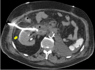
    Figure 1: Image shows right kidney with urinoma drained. Also showing
nephrostomy tube inserted.

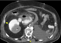
Figure 2: Large right urinoma on abdominal CT. Image taken before.
At postoperative day 20 he was readmitted for suspected sepsis after he presented with altered mental status. At presentation his serum creatinine was 9.4 and is potassium was 6.8. Abdominal CT showed a large retroperitoneal hematoma and a right perinephric fluid collection measuring 11 cm x 5.6 cm, and 17.6 cm craniocaudally (see Figure 1). Initial suspicion was that this fluid was likely an abscess, however IR drainage revealed 1200 ml of straw-colored fluid with analysis revealing creatinine content consistent with a urinoma (11mg/L). A drainage tube was placed (see Figure 2) with daily urine output of at least 1L. Patient’s renal function improved dramatically; creatinine was at baseline within 4 days of urinoma drainage and tube placement. Hemodialysis was discontinued at day 1 post procedure. A ureterogram showed no contrast extending beyond his mid ureter consistent with a ureteral obstruction. The drainage tube was then taken out and a nephrostomy tube placed. Plan was to further investigate his ureteral obstruction with eventual recanalization, but he opted for hospice care and was placed on comfort measures. He passed away 6 days post discharge. Serum creatinine at discharge was 0.8 mg/dl.
Discussion
Page phenomenon was first described in 1939 when Page wrapped canine kidneys in cellophane, the response measured was hypertension [2]. Since then, Page phenomenon or Page kidney have been described in the literature as extrinsic kidney compression, most commonly from a subcapsular hematoma. Page kidney from a subcapsular urinoma is rare. From our literature review, there have only been four reported cases as of 2009 [3]. The very first reported case was in 1984 by Patel, et al. [4]; it was a case involving a child born with posterior urethral valve. The other cases involved blunt force traumas and constrictive perinephritis [5,6]. The etiology in this case remains unclear as this patient did not have a history of direct renal instrumentation or trauma. It was theorized that his urinoma was likely related to his ureteral obstruction and/or complications from his surgery.
As earlier mentioned, a ureterogram was done with findings consistent with mid ureteral obstruction. Further investigation to determine the cause of obstruction was attempted but not done as patient opted for hospice. It was theorized, however, that this obstruction was likely due to an invasion by malignancy. Other possible causes, though considered unlikely, are ureteral stricture, ureteral stones and complications from his radical nephrectomy.
Most cases of Page kidney reported have been associated with hypertension. In the herein-presented case, while this patient was previously hypertensive from review of postoperative follow up records, he was normotensive at presentation on day 20 postoperative; this was believed to be due to sepsis and/or his being on multiple antihypertensive medications at presentation. He also presented with an acute kidney injury as defined by the KDIGO guidelines [7]. While there have been few reported cases of AKI resulting from Page kidney, none of them have involved a subcapsular urinoma making this a unique etiology of AKI.
Subcapsular urinoma should be suspected in patients with acute kidney injury who are high risk for Page kidney with or without hypertension. High risk patients include those with history of trauma, renal instrumentation (biopsy, surgery, ECSWL), renal cysts and malignancy. Such patients should be evaluated with imaging and treated with percutaneous drainage and tube placement if a diagnosis of subcapsular urinoma is made. Such patients should also undergo a ureterogram to rule out the presence of concomitant ureteral obstruction.
Compliance with Ethical Standards
Conflict of interest: The authors have declared that no conflict of interest exists.
Ethical approval: This article does not contain any studies with human participants or animals performed by any of the authors.
Informed consent: Informed consent was obtained from all individual participants included in the study.
References
- Dobson SJ, Jayakumar S, Velez, JCQ. Page Kidney as a Rare Cause of Hypertension: Case Report and Review of Literature. American Journal of Kidney Diseases. 2009; 54: 334-339.
- Page IH. The production of persistent arterial hypertension by cellophane perinephritis. JAMA. 1939; 113: 2046-2048.
- Takebayashi S, Matuzaki J. Page Kidney Caused by Progressive Perirenal Urinoma with Bridging Septa: Sonographic and CT Findings. European Journal of Radiology Extra. 2009; e29-e32.
- Patel MR, Mooppan MM, Kim H. Subcapsular urinoma: unusual form of “page kidney” in newborn. Urology. 1984; 23: 585-587.
- Matlaga BR, Veys JA, Jung F, Hutcheson JC. Subcapsular urinoma: an unusual form of page kidney in a high school wrestler. J Urol. 2002; 168: 672.
- Nomura S, Osawa G, Kinoshita H, Tanaka H. Page kidney with constrictive perinephritis. Nippon Jinzo Gakkai Shi. 1993; 35: 355-359.
- KIDIGO Clinical Practice Guidelines for Acute Kidney Injury 2012.