
Case Report
Austin J Clin Case Rep. 2020; 7(5): 1181.
Angle-Closure Glaucoma Secondary to an Iridociliary Cyst
Imane Chabbar*, Abdelkrim Boulanouar and Amina Berraho
Ophthalmology B, Ibn-Sina University Hospital, Morocco
*Corresponding author: Imane Chabbar, Ophthalmology B, Ibn-Sina University Hospital, Morocco
Received: October 27, 2020; Accepted: November 10, 2020; Published: November 17, 2020
Abstract
Iridociliary cysts are relatively uncommon. They are asymptomatic in the majority of cases. However, they can cause angle-closure glaucoma by repelling the structures of the angle. UBM is the examination of choice to study the mechanism of angle-closure glaucoma secondary to an iridociliary cyst and to differentiate it from melanoma and solid tumors.
Case Presentation: A 49-year-old female referred to the university hospital for evaluation and management of a chronic angle-closure glaucoma resistant to treatment. On examination, visual acuity was 10/10 in OD and 1/10 in OS, a reduced anterior chamber in both eyes. The intraocular pressure was 19 mmhg in the right eye and 24 mmhg in the left eye under triple hypotonic therapy. Gonioscopy demonstrated a closed angle with anterior insertion of the iris. UBM shows shallow anterior chambers with closed angles and a plateau iris configuration in both eyes, and in the left eye a ciliary body cyst.
Conclusion: Ultrasound biomicroscopy is a valuable technique in diagnosing iridociliary cysts.
Keywords: Iridociliary Cyst; Angle-Closure Glaucoma; Ultrasound Biomicroscopy
Introduction
Iridociliary cysts are relatively uncommon. They can be of primitive or secondary origin [1]. They are asymptomatic in the majority of cases. However, they can increase in volume and involve secondary glaucoma, corneal endothelial decompensation and show a fluid-debris level reminiscent of a pseudohypopyon [2]. Ultrasound Biomicroscopy (UBM) is a useful option that completes clinical examination. It can provide a high-resolution view of the iris and ciliary body and distinguish primary cysts from solid uveal tumors [3].
The objective of this work is to underline the interest of UBM in the evaluation of angle-closure glaucoma secondary to an irido-ciliary cyst.
Methods
We report an observation of a patient presenting a chronic angle-closure glaucoma not stabilized under medical treatment, referred to the university hospital for compete evaluation and adequate management. The patient benefited from a careful interrogation, a complete ophthalmological examination with additional investigations: visual fields, UBM, anterior segment and optic nerve OCT.
Case Presentation
A 49-year-old woman, without pathological history, treated for chronic bilateral and glaucoma since 2008 with triple therapy. The ophthalmological examination found visual acuity in OD 10/10 (+2,00) and in OS 1/10 (+3,00), a clear cornea with an anterior chamber reduced in periphery in both eyes. The intraocular pressure was 19 mmhg in the right eye and 24 mmhg in the left eye under two hypotonic eye drops: Duotrav* (Travoprost - Timolol) + Alphagan* (Brimonidine). Gonioscopy demonstrated a grade 0 to1 angles 360º bilaterally with anterior insertion of the iris. The eye fundus examination (Figure 1a, 1b) shows pathological papillary excavation with C/D at 4/10 in OD and 8/10 in OS.
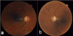
    Figure 1: Eye fundus examination showing a pathological papillary excavation
with C/D at 4/10 in OD (a) and 8/10 in OS (b).
UBM (Figure 2) shows shallow anterior chambers with narrow angles and a plateau iris configuration in both eyes, and in the left eye a ciliary body cyst.
Anterior segment OCT (Figure 3) demonstrated narrow angles consistent with gonioscopy and UBM findings, but not showing the ciliary body cyst.
Discussion
Iridociliary cysts can be either of primary origin from the pigment epithelium more rarely from the iris stroma, or secondary to trauma or surgery or prolonged myotics use [1,2]. Iridociliary cysts are a rare etiology for plateau iris syndrome, and they are characterized by a convex configuration of the peripheral iris that leads to plateau iris configuration and a narrow angle. These cysts can cause angle-closure glaucoma by repelling the structures of the angle [4].
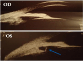
    Figure 2: UBM showing shallow anterior chambers with narrow angles
bilaterally and a ciliary body cyst in the left eye (arrow).
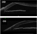
    Figure 3: Anterior segment OCT showing narrow angles bilaterally but not
showing the ciliary body cyst.
UBM is the examination of choice for elucidating the mechanism of angle-closure glaucoma secondary to an iridociliary cyst by showing a regular cystic formation thin walled with echogenic content. It makes it possible to appreciate the impact on the angle and decide on the appropriate therapeutic approach [5]. UBM also allows the differential diagnosis of iris melanoma and cysts of the iris pigment epithelium. Iris and ciliary body cysts are often mistaken as a mass or tumor; most commonly an iris melanoma [6]. UBM is a simple diagnostic procedure that can help in establishing the correct diagnosis and thereby avoid unnecessary and costly procedures.
Concerning the treatment of iridociliary cysts, Argon laser iridoplasty [7] makes it possible to oppose the angle closure and to free access of aqueous humor to the trabeculum. Peripheral iridectomy is ineffective because it does not necessarily target the cyst. Yag laser irido-cystectomy [8] can be used, but the risk of recurrence after healing is very possible. Finally, the filtration surgery is indicated if glaucoma remains uncontrolled.
Conclusion
Iridociliary cysts are benign cystic lesions that can compromise the angle. UBM represents a valuable technique in diagnosing iridociliary cysts and studying the anterior segment structures, particularly in glaucomatous pathology. In our case, UBM allowed us to diagnose angle-closure glaucoma secondary to an iridociliary cyst and to follow its evolution.
References
- Shields CL, Kancherla S, Patel J, Vijayvargiya P, Suriano MM, Kolbus E, et al. Clinical survey of 3680 iris tumors based on patient age at presentation. Ophthalmology. 2012; 119: 407-414.
- Shields JA. Primary cysts of the iris. Trans Am Ophthalmol Soc. 1981; 79: 771-809.
- Marigo FA, Esaki K, Finger PT, H Ishikawa, D S Greenfield, J M Liebmann, et al. Differential diagnosis of anterior segment cysts by ultrasound biomicroscopy. Ophthalmology. 1999; 106: 2131-2135.
- Thomas R, Mulligan N, Aylward GW, Billson FA. Angle closure glaucoma due to iris and ciliary body cysts. Aust N Z J Ophthalmol. 1989; 17: 317-319.
- Kunimatsu, Shiho, Araie, Makoto, Ohara, Kunitoshi, Hamada, Chikuma. Ultrasound biomicroscopy of ciliary body cysts. Am. J. Ophthalmol. 1999; 127: 48-55.
- McWhae JA, Rinke M, CrichtonAC, Van Wyngaarden C. Multiple bilateral iridociliary cysts: ultrasound biomicroscopy and clinical characteristics. Can. J. Ophthalmol. 2007; 42: 268-271.
- Crowston JG, Medeiros FA, Mosaed S, Weinreb RN. Argon laser iridoplasty in the treatment of plateau-like iris configuration as result of numerous ciliary body cysts. Am J Ophthalmol. 2005; 139: 381-383.
- Kuchenbecker J, Motschmann M, Schmitz K, Behrens-Baumann W. Laser iridocystotomy for bilateral acute angleclosure glaucoma secondary to iris cysts. Am J Ophthalmol. 2000; 129: 391-393.