Case Report
Austin J Clin Case Rep. 2020; 7(6): 1188.
Early Onset Necrotizing Enterocolitis (NEC) in Twin Pregnancy: A Case Report
Wegdan Helmy Mawlana1,2*, Asmaa Osman2 and Atallah Al Howeiti2
1Department of Pediatrics and Neonatology, Tanta University Hospital, Egypt
2Divison of Neonatology, Department of Pediatrics, King Salman Armed Forces Hospital, Saudi Arabia
*Corresponding author: Wegdan Helmy Mawlana, Associate Professor of Pediatrics and Neonatology, University Hospital, Egypt
Received: December 09, 2020; Accepted: December 24, 2020; Published: December 31, 2020
Abstract
Early onset necrotising enterocolitis developed on the third day of life in twin infants born at 34 week gestation. Both were on mixed breast milk and term formula. Both infants developed bloody stool with abdominal distension. Radiological finding of NEC were evident on serial x-rays. Sepsis work up was negative. Baby boy was managed conservatively and discharged home. Unfortunately baby girl had aggressive NEC that mandate laparotomy with multiple areas resected and long term complications developed.
Keywords: Twin; Necrotizing Enterocolitis; Preterm Infants
Introduction
Necrotizing Enterocolitis (NEC) is a devastating gastrointestinal problem that could progress to serious complications. It is mainly encountered in the premature neonate (with birth weight <1500 gram) [1]. Approximately 10% of NEC cases may be presented in term infants. While NEC in preterm infants usually present in the third week of life, NEC in term infants commonly present by the first week of life [2]. NEC is common amongest twins, and it is always in the first born of the twins [3]. We reported a case of NEC in both twin born at 34 weeks gestational age.
Case Presentation
A dichorionic diamniotic twin boy and girl were born to unbooked 38-year-old Saudi female, Gravida 6 para 5. Her serology was nonreactive. Her medical history was unremarkable. Both babies were delivered by spontaneous vaginal delivery. They were born vigorous, required only initial steps of resuscitation. They had Apgar score 7 and 8 at 1, and 5 min respectively. Twin A baby girl had birth weight of 1.90 kg (10-50% percentile), length 44 cm (50% tile) and Head Circumference (HC) 33 cm (50-90% percentile). Twin B (baby boy) had birth weight of 1.93 kg, (10-50% percentile), length 44 cm (50% percentile), and HC of 32 cm (50-90% tile). Both had mild respiratory distress with mild intercostal and subcostal retraction so they were admitted to NICU. Both babies were active, not dysmorphic and had mild respiratory distress needed nasal CPAP for few hours and weaned to room air by second day of life. Blood culture was taken at birth and started empirical antibiotics (Ampicillin and Gentamycin).
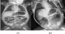
Twin A (baby girl): Feeding was started gradually with Expressed Breast Milk (EBM) but the mother was unable to provide enough EBM so term formula was introduced. Blood culture showed no growth for 48 hours and discontinued antibiotics. On the third day of life, she developed recurrent bradycardia and desaturation together with abdominal distension, grunting and bleeding per rectum. Baby was intubated and connected to mechanical ventilator, septic work up was done. Abdominal x-ray showed extensive pneumatosisintestinalis (Figure 1). Antibiotics were restarted again (vancomycin, meropenem and Flagyl) for NEC (Bell stage II). Laboratory finding summarized in (Table 1). Pediatric surgery consultation done and advised for conservative management. On day 8 of life, x-ray showed pneumoperitoneum. Laparotomy was done and showed multiple patches of gangrene in the entire jejunum and ileum then jejunostomy was performed. About 30 cm of jejunum were resected. Ileocecal valve was preserved. She was kept NPO for 14 days and on triple antibiotics. Trophic EBM feeding was reintroduced by day 14 post operation and gradually increased according to the local feeding protocol. Baby continued to have high stoma output. Pediatric gastroenterology recommended shifting to hydrolysed formula (Neocate). However, baby continued to have high stoma output. On day 59 of life, baby underwent her second operation for closure of the stoma. Feeding was restarted gradually with intermittent abdominal distension that mandated contrast study which was not conclusive. On day 75 of life, baby did not tolerate further progress of feeding and developed greenish vomiting. Baby underwent her third operation with exploration laparotomy. Multiple areas of adhesions and strictures were found all over the intestine so adhesion lysis was done together with resection of 4 cm of the ileum, ileocecal valve and appendix and 3 cm of the ascending colon with end to end anastomosis. After that baby was kept NPO for another 10 days, feeding proceeded slowly with no issues. Because of this stormy course with all complications of central lines associated infection and parenteral nutrition (PN) - associated liver disease. Baby was discharged home at the age of 3 months with weight of 3.7 kg (<3%). She still has frequent episodes of loose stool with electrolytes disturbance and poor weight gain with close follow up by multidisciplinary team of gastroenterology, dietitian and pediatric surgery.

    Figure 1: (A1): Abdominal X-ray for twin A show pneumatosis intestinalis
and intrahepatic portal venous gas, (A2): Abdominal X-ray for twin A show
pneumoperitoneum.
Investigations
Twin A
Twin B
Hb (%)
15.7
20.6
Haematocrit
43.6
54.8
WBC
1.91
2.29
Differential count (%)
Lymph-29; MONO- 14; EOS-7
LYMPH-28; MONO-20; EOS-0
Platelet count
50
200
CRP
1
1
Clotting Studies:
PT
PTT
INR
12.6
32.9
0.97
14.7
35.9
1.12Total proteins (gm/dL)
40
39
Albumin (gm/dL)
19
22
Aspartate aminotransferase (AST)(U/L)
238+
24
Alanine aminotransferase (ALT)
38
14
Blood group
B+
B+
Cultures (blood/urine)
Negative
Negative
Table 1: Laboratory investigation in both infants at the onset of NEC.
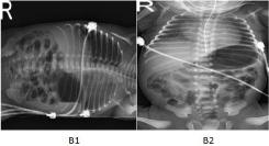
Twin B (baby boy): Feeding was started on term formula on day 1 and adlib feed established by the second day of life. Interestingly, baby had frank blood in stool on day 3 same as his sister but the abdomen was soft, not distended, non-tender with active bowel sounds. Abdominal x-ray showed demonstrated pneumatosis intestinalis (Figure 2). Investigations performed in twin B are showing in Table 1. Twin B received the same management as his sister being kept NPO, and triple antibiotics for 10 days. Blood culture came back negative. Feeding was restarted with exclusive breast milk, gradually reach full feeds. Baby discharged home on breast milk with appropriate weight gain with discharge weight of 2.7 kg (10%) on day 37 of life followed by dietitian and gastroenterology.
Discussion
Surgical NEC in twin pregnancy had been reported in 20% of babies who developed NEC in the study by Burjonrappa et al 2014. Seven of the twin pregnancies were dizygotic. It could be explained that preterm twin deliveries do have a higher incidence of NEC than singleton preterm delivery [4]. NEC in twin pregnancy neonates showed a female preponderance compared to singletons and occurred universally in the first born of the twins [5]. Not surprising that the first born was female that had aggressive course. This preponderance of NEC in the first born of the two twins also suggests that hormonal changes at the time of delivery affect the first born more than the second born twin may have a role in the development of NEC [6]. Our twins were dizygotic twin. This discordance in the development of NEC has been noted in other twin studies too. Although dizygotic twin have different immune system but they have many shared genes [4]. While the median age of neonates developing NEC is usually around three weeks in the singleton and twin pregnancies, our twin developed NEC too early by the third day of life.

    Figure 2: (B1): Lateral decubitus X-ray shows pneumatosis intestinalis. No
perforation, (B2): Abdominal X-ray showspneumatosis intestinalis.
The presentation of NEC is variable, from mild course require only conservative management as the case in twin II to an aggressive form requiring surgical laparotomy as in twin I.
Sepsis contributes significantly to NEC specially gram negative bacteria, however, blood culture was negative in both twins excluding infection as underlying cause for NEC in our case. While our twin received breast milk in their first feed, term formula was introduced after that due to insufficient breast milk. Previous studies have shown that human milk reduces the risk of NEC due to its content of many factors that may not be present in formula milk such as leukocytes, oligosaccharides, immunoglobulins such as IgA, lactoferrin, and many others that could provide antimicrobial and anti-inflammatory action [7,8]. Afzal et al reported an interesting case report of almost similar scenario as our cases of 34 weeks twin who developed bloody stool on the third day of life. Bloody stools in healthy appearing infants are most commonly due to allergic colitis from food allergy as in Afzal et al case report [9]. He hypothesized that milk protein allergy would be the most probable diagnosis for his case due to inutero maternal sensitization to cow milk and the antigen crossing the placenta could trigger allergic response on the gut. Although cow milk allergy could be considered in the differential diagnosis of our case, mother history was unremarkable. Biopsy of the resected segment in twin I showed necrotizing enterocolitis as the underlying cause of bloody stool. Breast milk was reintroduced in Twin II after 10 days of Nil Per Mouth (NPO) with no recurrence of bloody stool and baby was discharged with average weight gain. This also abolished the diagnosis of cow milk allergy as an underlying cause for our case.
The radiologic findings of NEC are not specific; pneumatosis intestinalis could also result from any other causes of bowel ischemia as volvulus or sepsis which makes the diagnosis of NEC is challenging [10]. The evaluation of bowel by ultrasound has been shown to have high specificity for bowel necrosis [11]. Unfortunately our twin was not evaluated by bowel U/S was due to lack of expertise in our center.
References
- Hackam D, and Caplan M. Necrotizing enterocolitis: Pathophysiology from a historical context. Semin Pediatr Surg. 2018; 27: 11-18.
- Bazacliu C, Neu J. Necrotizing Enterocolitis: Long Term Complications. Curr Pediatr Rev. 2019; 15: 115-124.
- Meister AL, Doheny KK, Travagli RA. Necrotizing enterocolitis: It's not all in the gut. Exp Biol Med (Maywood). 2020; 245: 85-95.
- Burjonrappa S, Shea B, Goorah D. NEC in Twin Pregnancies: Incidence and Outcomes. Journal of Neonatal Surgery. 2014; 3: 45.
- Stewart CJ, Marrs EC, Nelson A, Lanyon C, Perry JD, Embleton ND, et al. Development of the preterm gut microbiome in twins at risk of necrotising enterocolitis and sepsis. JEPLoS One. 2013; 8: e73465.
- Chauhan SP, Scardo JA, Hayes E, Abuhamad AZ, Berghella V. Twins prevalence, problems and preterm birth. Am J Obstet Gynecol. 2010; 10: 310-315.
- Sullivan S, Schanler RJ, Kim JH, Patel AL, Kiechl-Kohlendorfer U, Chan GM, et al. An exclusively human milk-based diet is associated with a lower rate of necrotizing enterocolitis than a diet of human milk and bovine milk-based products. J Pediatr. 2010; 156: 562-571.
- Good M, Sodhi CP, Egan CE, Amin Afrazi, Hongpeng Jia, Yukihiro Yamaguchi, et al. Breast milk protects against the development of necrotizing enterocolitis through inhibition of Toll-like receptor 4 in the intestinal epithelium via activation of the epidermal growth factor receptor. Mucosal Immunol. 2015; 8: 1166-1179.
- Afzal B, Elberson V, McLaughlin C, Kuma VH. Early onset necrotizing enterocolitis (NEC) in premature twins .J Neonatal Perinatal Med. 2017; 10: 109-112.
- Meister AL, Doheny KK, Travagli RA. Necrotizing enterocolitis: It's not all in the gut Exp Biol Med (Maywood). 2020; 245: 85-95.
- Kim JH. Role of Abdominal US in Diagnosis of NEC. Clin Perinatol. 2019; 46: 119-127.