Case Report
Austin J Clin Case Rep. 2021; 8(4): 1205.
Gamma Knife for Recurrent Ameloblastoma with Cavernous Sinus Metastasis: A Case Report
He B, Yang S and Li D*
Department of Radiation Oncology, Third Medical Center of Chinese PLA General Hospital, Beijing, China
*Corresponding author: Dongbo Li, Department of Radiation Oncology, Third Medical Center of Chinese PLA General Hospital, 69 Yongding Road, Haidian District, Beijing, China
Received: March 31, 2021; Accepted: April 16, 2021; Published: April 23, 2021
Abstract
Ameloblastoma (AME) is a rare, benign intraosseous progressively growing odontogenic tumor. Due to its invasive behavior, the rate of recurrence is high. Recurrent AME tends to transform malignantly and metastatic. Lung is the most common sites of AME metastasis, followed by lymph nodes. Here we present a case of AME with intracranial metastasis. A 26-year-old woman who had recurrent AME in the left jaw. After the second resection, AME metastasis to the cavernous sinus, sellar and suprasellar regions. Because the metastatic tumor was unresectable, she received Gamma Knife instead. After 3 years follow-up, the tumor was well controlled. In conclusion, Gamma Knife can be a feasible option for unresectable Oligometastatic AME.
Keywords: Ameloblastoma; Cavernous sinus metastasis; Gamma knife
Introduction
Ameloblastoma (AME) is one of the most common tumor arise from epithelial dental lamina. Luo et al. reported on 1309 benign oral tumors in China, of which AME was the second most common (36.52%) [1]. AME was first described in 1827 by Cusack [2], and known for its aggressive behavior and high recurrence rate. The 2017 WHO classification identifies AME as a benign intraosseous progressively growing epithelial odontogenic neoplasm. Which characterized by expansion and a tendency or local recurrence if no adequately removed [3].
About 2% of ameloblastomas will develop metastases. Most of AMEs metastatic to the lung (70-85 % of cases), followed by lymph nodes (20% of cases) [4]. Although metastases of AME have a benign histologic appearance. Yet, 30% of the patients will die in five years [5]. The reason for such a low survival rate is the lack of effective drug. As a local treatment, radiotherapy has limited effect on metastatic tumor. However, for oligometastatic disease, especially the unresectable one, radiotherapy can remedy the deficiency of drug therapy.
Herein, we present a case of Oligometastatic AME. The unresectable metastatic AME was well controlled with Gamma knife.
Case Presentation
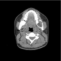
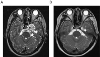
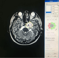
A 26-year-old woman with AME had a recurrent exophytic tumor over the left jaw for more than 6 years. Initially, she had noticed a painless, swollen mass on her jaw. However, the mass grew over the course of approximately one year (Figure 1). She visited a local outpatient hospital where a biopsy showed AME (Figure 2A and 2B) and she underwent tumor curettement. Three years after the first surgery, the tumor recurred in the left jaw. Extensive resection of the tumor was performed. One year after the second surgery, the patient returned to the hospital. She presented with double vision, disordered left eye movement, and left eyelid ptosis. Magnetic Resonance Imaging (MRI) showed the tumor metastasized to the cavernous sinus, sellar and suprasellar regions (Figure 3). Biopsy was performed, and pathology showed AME again (Figure 4). Because the tumor was unresectable. She received Gamma Knife with a peripheral dose of 11Gy and a central dose of 24Gy (45% isodose curve) in 1 fraction. MRI was reviewed 3 months after the Gamma Knife (Figure 4); the tumor was significantly reduced (Figure 5). The patient’s symptoms were markedly resolved. Her AME remained well-controlled 3 years following Gamma Knife.

    Figure 1: CT of mandible show cystic bone destruction in the molar area. The
feature is consistent with AME.

    Figure 2: Microscopic analysis of AME. A) Microscopy shows many small,
discrete tumor islands. This feature is consistent with AME (hematoxylineosin
stain, magnification ×100). B) Hyperchromatism and increased mitotic
index were observed (hematoxylin-eosin stain, magnification × 400).

    Figure 3: T2-weighted MRI of the cavernous sinus. A) The tumor is seen in
the left sphenoid bone with invasion of the cavernous sinus. B) The tumor as
seen three months after the completion of radiotherapy (white stars).

    Figure 4: Microscopic analysis of cavernous sinus tissue. A) Microscopy
shows many small, discrete tumor islands. This feature is consistent with
AME (hematoxylin-eosin stain, magnification × 100). B) Hyperchromatism
and increased mitotic index were observed (hematoxylin-eosin stain,
magnification × 400).

Figure 5: The Gamma Knife plan and isodose distribution curves.
Discussion
Metastasizing ameloblastoma is extremely rare, and there is currently no established treatment protocol. Although the 2017 WHO classified metastatic AME as benign tumor. However, due to the lack of effective drug, uncontrolled metastasis is still the main cause of AME death [3].
In all kinds of metastatic sites, intracranial ameloblastomas are particular rear. A study by Olaitan et al. showed only 3 patients (<1%) get intracranial metastasis in the large population of 315 ameloblastoma patients [6]. Unlike AME lung or lymph node metastasis, most of AME intracranial metastases are oligometastases. Besides, intracranial metastases are often difficult to completely remove. Even complete resections always accompanied by cranial nerve injury. A lot of clinical studies show improved survival when radical local therapy added to standard systemic therapy for oligometastatic disease [7]. Moreover, radiotherapy especially stereotactic radiotherapy has advantages over neurosurgery in terms of neuroprotection. Due to the lack of drug therapy. Radiotherapy for metastatic AME become more important.
In this case, our patient was a young woman gone through two mandible AME reaction, and got cavernous sinus metastasis. The left abducent nerve and oculomotor nerve were compressed by the tumor. She presented with double vision, disordered left eye movement, and left eyelid ptosis. Since the tumor was unresectable, we suggested her taking radiotherapy. After fully explaining the possible risks and sequelae of developing radiation-induced malignancies. She consented to take Gamma Knife for her intracranial metastases. Due to the rarity of intracranial AME, There is no standardized guideline for her treatment with radiotherapy [8-11]. Considering the tolerance of cranial nerve and according to the experience of our department. We give her Gamma Knife with a peripheral dose of 11Gy and a central dose of 24Gy (45% isodose curve) in 1 fraction. Symptoms improved significantly after treatment. Unresectable AME was successfully salvaged by Gamma Knife. Without any medication the metastatic AME was well-controlled after 3 years of follow-up. The patient’s neurological function is fine. And there is no serious side effect of radiotherapy.
Gamma Knife for intracranial metastatic AME was safe and effective for our patient. We will continue to follow up.
Conclusion
We suggest Gamma Knife may be an effective option for intracranial metastatic AME. Peripheral dose of 11Gy and a central dose of 24Gy (45% isodose curve) in 1 fraction is a good dose reference. For oligometastatic AME, Radiotherapy can remedy the deficiency of drug therapy.
Acknowledgement
We appreciated Xingyu He for Provide pathological section for the manuscript.
Availability of Data and Materials
All data generated or analyzed during this study are included in this published article.
Authors’ Contributions
Dongbo Li (Corresponding author): Carried out contouring the patient, drafted, generally evaluated and write the manuscript, Bo He: Carried out the patient interrogation and follow-up, write the manuscript, Shuming Yang: Evaluated the patient’s radiographs and made calculations. All authors read and approved the final manuscript.
Patient Consent for Publication
Written informed consent for publication of their clinical details and clinical images was obtained from the patient. A copy of the consent form is available for review by the Editor of this journal.
References
- Luo HY, Li TJ. Odontogenic tumors: A study of 1309 cases in a Chinese population. Oral Oncol. 2009; 45: 706-711.
- Cusack JW. Report of the amputations of the lower jaw. Dublin Hosp Rec. 1827; 4: 1-38.
- El-Naggar AK, Chan JKC, Grandis JR, Takata T, Slootweg PJ. Eds. WHO Classification of Head and Neck Tumours. 4th ed. Lyon, France. IARC. 2017.
- Jayaraj G, Sherlin HJ, Ramani P, Premkumar P, Natesan A, Ramasubramanian, et al. Metastasizing Ameloblastoma - A perennial pathological enigma? Report of a case and review of literature. J of Cranio Maxillofac Surg. 2014; 42: 772-779.
- Peter R, James J, Sciubba DMD, Hans PP. Commentary Splitters or lumpers: The 2017 WHO Classification of Head and Neck Tumours. J of the American Dental Association. 2018; 149: 567-571.
- Olaitan AA, Adeola DS, Adekeye EO. Ameloblastoma: clinical features and management of 315 cases from Kaduna, Nigeria. J Craniomaxillofac Surg. 1993; 21: 351-355.
- Guckenberger M, Lievens Y, Bouma AB, Collette L, Dekker A, deSouza NM, et al. Characterization and classification of oligometastatic disease: a European Society for Radiotherapy and Oncology and European Organization for Research and Treatment of Cancer consensus recommendation. Lancet Oncol. 2020; 21: e18-e28.
- Chehal A, Lobo R, Naim A, Azinovic I. Ameloblastoma of the maxillary sinus treated with radiation therapy. Pan Afr Med J. 2017; 26: 169.
- Atkinson CH, Harwood AR, Cumings BJ. Ameloblastoma of the jaw. A reappraisal of the role of megavoltage irradiation. Cancer. 1984; 53: 869-873.
- Philip M, Morris CG, Werning JW, Mendenhall WM. Radiotherapy in the treatment of ameloblastoma and ameloblastic carcinoma. J Hong Kong Coll Radiol. 2005; 8: 157-161.
- Kennedy WR, Werning JW, Kaye FJ, Mendenhall WM. Treatment of ameloblastoma and ameloblastic carcinoma with radiotherapy. Eur Arch Oto Rhino Laryngol. 2016; 273: 3293-3297.