Clinical Image
Austin J Clin Case Rep. 2021; 8(5): 1212.
Not a Huge but Two Tumors
Jie He*
Department of Thoracic Surgery, National Cancer Center/ National Clinical Research Center for Cancer/Cancer Hospital, China
*Corresponding author: Jie He, Department of Thoracic Surgery, National Cancer Center/National Clinical Research Center for Cancer/Cancer Hospital, Chinese Academy of Medical Sciences and Peking Union Medical College, Panjiayuan Nanli No 17, Beijing, 100021, People’s Republic of China
Received: April 28, 2021; Accepted: May 20, 2021; Published: May 27, 2021
Clinical Image
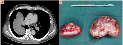
A 73-year-old man presented to the thoracic surgery clinic with progressive difficulty breathing for about 3 months. He had a benign spindle cell tumor (about 3 centimeter in diameter) in right lung upper lobe, which had been resected by thoracotomy surgery 15 years earlier. The physical examination showed weakened right respiratory sounds. Computed tomography of the chest revealed a huge mass closed to the pulmonary vessels and bronchus, which measured more than 13 cm in greatest dimension (Panel A). Thoracotomy surgery was performed for the tumor resection again. Two separated tumors, instead of one huge tumor, originated from visceral pleura were found with well encapsulated (Panel B). And the histopathological analysis revealed solitary fibrous tumor. The immunohistochemical analysis was positive for Desmin-protein and CD34. Just followingup was initiated after surgery, and the patient was doing well after 3 months.
