
Case Report
Austin J Clin Case Rep. 2022; 9(1): 1241.
Female Genital Schistosomiasis, a Neglected Differential of Cervical Precancerous and Cancerous Lesion: a Wakeup Call for on-Job Training for Healthcare Workers in Endemic Areas
Kaizilege GK¹, Kiritta R¹*, Chuma C¹, Ndaboine E¹, Ottoman O², Elias E², Zinga MM³ and Mazigo HD³
¹Department of Obstetrics and Gynecology, Weill Bugando School of Medicine, Catholic University of Healthy and Allied Sciences, Mwanza, Tanzania
²Department of Pathology, Weill Bugando School of Medicine, Catholic University of Healthy and Allied Sciences, Mwanza, Tanzania
³Department of Parasitology, Weill Bugando School of Medicine, Catholic University of Healthy and Allied Sciences, Mwanza, Tanzania
*Corresponding author: Richard Kiritta, Department of Obstetrics and Gynecology, Weill Bugando School of Medicine, Catholic University of Healthy and Allied Sciences, P.O. Box 1464, Mwanza, Tanzania
Received: February 01, 2022; Accepted: February 25, 2022; Published: March 04, 2022
Abstract
Background: Female genital schistosomiasis is a gynecological disease caused by Schistosoma haematobium. The uterine cervix appears to be the most affected area with most cases found in sub-Saharan Africa. Though both males and females can be affected, the consequences of genital schistosomiasis are more pronounced in females with reported increased risk of HIV transmission and malignancy of female genitalia. Female genital schistosomiasis of the cervix shares similarities in presentation to those of cervical intraepithelial neoplasm and sexual transmitted infections, making diagnosis challenging and requiring high degree of suspicion.
Case Presentation: We report three cases of African women referred to our facility with a presumptive diagnosis of cervical precancerous/cancerous lesion. Initial workout at our facility was in keeping with early stages of cervical precancerous/cancerous lesion that necessitated excision as per standardized protocol. Histopathological analysis of excised tissue revealed schistosomiasis of the cervix.
Conclusion: Schistosomiasis of the cervix shares similarities with cervical precancerous/cancerous lesions and should be suspected in women presenting with chronic inflammatory conditions of the cervix or features suggestive of precancerous/cancerous cervical lesions. Screening for female genital schistosomiasis should be incorporated in an already existing screening protocol for cervical cancer and sexual transmitted diseases.
Keywords: Genital schistosomiasis; Cervical cancer; Schistosoma; Vaginal discharge
Introduction
Schistosomiasis is an acute and chronic neglected tropical disease caused by parasitic flatworms belonging to the genus Schistosoma. In the year 2019, it was estimated that approximately 240 million people were affected with the disease globally [1] and around 659 million people were vulnerable to contract the disease. It is highly prevalent in tropical and subtropical regions with 95% of the disease burden occur in sub-Saharan Africa [2]. Contact with infested water bodies during daily activities such agriculture, fishing, swimming and washing clothes/utensil predisposes individuals to Schistosoma infection. Inadequate hygiene and contact with infected water make children especially vulnerable to infection [1].
Approximately 56 million African women and young girls are infected with schistosomiasis [3]. The disease especially caused by S. haematobium does not only affect the urinary tract but also invade the female reproductive tract leading into detrimental effects to the organs, this is referred to as female genital schistosomiasis [2,4]. Despite the fact that schistosomiasis can invade the entire female reproductive tract, the uterine cervix is the most common affected site [5,6].
Clinical presentation of cervical schistosomiasis shares similarities to that of precancerous/cancerous cervical lesions and sexual transmitted infections, making the diagnosis challenging in the absence of a high degree of suspicion for this neglected tropical disease. Three cases of FGS with prior presumptive diagnosis of cervical precancerous/cancerous lesions are hereby discussed.
Case Presentation
Case 1
37 years old female referred from a peripheral regional hospital suspected to have cervical cancer, presented at our facility with longstanding lower abdominal pain, abnormal vaginal bleeding mostly post-coital and copious vaginal discharge. She is Para 9, known patient with HIV on ART for 11 years now. She suffered from schistosomiasis (urinating blood) at the age 8 years in 1992 which resolved after using herbal medication. Her review of other system, obstetrics and gynecology history was uneventful.
Gynecological examination for cervical cancer screening was done and revealed normal vulva and vaginal walls. There was non-foul smelling copious vaginal discharge, cervix had a normal morphology but hyperemic on the whole ectocervix. Visual inspection by application of acetic acid produced aceto-white reaction on the cervix and an impression of precancerous cervical lesion was reached which necessitated loop electrosurgical excision procedure (LEEP) as an option of treating precancerous cervical lesion. The excised tissue samples were taken for histopathological analysis which revealed multiple eggs of Schistosoma haematobium associated with mixed inflammatory cells, mainly eosinophils as shown in Figure 1 below.
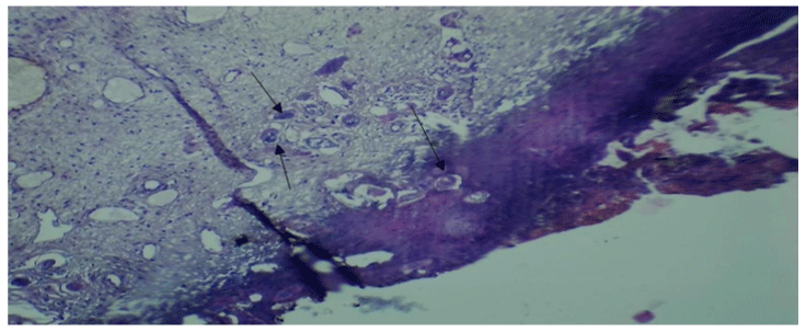
    Figure 1: Tissue section from the ectocervix showing multiple accumulations

of active schistosoma eggs (pointed by black arrows) surrounded with

inflammatory cells (x10hpf).
Case 2
A 35 years old female P2L2 HIV negative was referred to our facility from a peripheral cervical cancer screening clinic for LEEP as she was diagnosed to have large aceto-white lesion following application of acetic acid during cervical cancer screening. She reported history of lower abdominal pain, painful coitus, abnormal vaginal discharge and contact vaginal bleeding for more than 5 months. Her last delivery was eight years ago, she reports failure of conception for three years despite regular coitus. No surgical history and her past medical, obstetric, allergy and drug history were uneventful.
She was clinically stable and pelvic examination revealed normal vulva and vaginal walls, normal sized cervix, acetic acid was applied and large aceto-white lesion were seen extending to the posterior vaginal fornix. Loop electrosurgical excision procedure was done with an intention of treating cervical precancerous lesion. Cervical tissues were taken for histopathology and revealed chronic granulomatous inflammation with multiple calcified Schistosoma haematobium eggs, multinucleated giant cells, epithelioid cells, lymphocytes and numerous multiple eosinophils as shown in Figure 2 below.
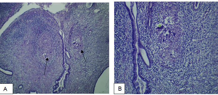
    Figure 2: Tissue section from the endocervix showing multiple granuloma

lesion (pointed in black arrow) (x4 hpf in figure a) and calcified Schistosoma

eggs (pointed with green arrows) surrounded with inflammatory cells. (x10

hpf in figure b).
Case 3
24 years old female P0+2, with history of foul-smelling vaginal discharge, lower abdominal pain and abnormal vaginal bleeding. Presented at our screening unit for cervical cancer screening after being treated for sexual transmitted infections (STI) several time with remission. She had received treatment for STI twice without improvement. She was married, doing small business, and had history of two spontaneous abortion. She had no history of chronic illness or any major/minor surgery, her menstrual flow was normal.
Her physical examination was uneventful, pelvic examination revealed a normal vulva and vagina wall with foul smell discharge, the cervix had ulcer like lesion at 6’o clock which was bled on contact. We had an impression of cervical cancer and tissue biopsy was taken at the site of lesion for histopathological examination. At examination, numerous accumulations of both active and calcified Schistosoma eggs surrounded with numerous multiple eosinophils, lymphocytes and neutrophils were observed (Figure 3).
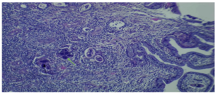
    Figure 3: Tissue section from the endocervix showing accumulation of both

active Schistosoma eggs (pointed by green arrow) and calcified Schistosoma

eggs (pointed by black arrow) surrounded with inflammatory cells (x10hpf).
Discussion
Tanzania ranks second among African countries with high prevalence of Schistosomiasis with reported prevalence range from 51.5% to 100%, S. haematobium (urogenital) and S mansoni (intestinal) being the commonest species [7-9].
Despite a high reported prevalence of female genital schistosomiasis and a well-known reproductive and social complication associated with female genital schistosomiasis in Tanzania, the disease is among the neglected conditions leading to underdiagnosis and mistreatment [5,6].
S. haematobium has often been associated with urogenital schistosomiasis. Mature/adult schistosomes are capable of leaving the portal vein and colonize the peri vesical plexus resulting in infection to the urinary bladder and surrounding reproductive organs in both female and male [10]. In female, eggs released from the peri vesical plexus migrates to genital organs leading to chronic granulomatous inflammatory lesions in the ovaries, fallopian tubes, cervix, vagina, and vulva thus causing female genital schistosomiasis [5,11].
Female genital schistosomiasis has been associated with significant morbidity and may cause complications such as chronic pelvic pain, infertility, spontaneous abortion or ectopic pregnancy, stress urinary incontinency and genital ulcers which may increase a woman’s risk for acquisition and transmission of HIV [4,5,12-14]. As was seen in our cases, one of our patients had history of two first trimester spontaneous abortion, the other had secondary infertility as she had not conceived in the past 5-years despites several efforts done with regard to conception while one patient was HIV positive. Even though the cause of the above-mentioned conditions cannot be sorely attributed to schistosomiasis in this report, it is reasonable to suspect schistosomiasis related complications as a possibility.
Female genital schistosomiasis may present with dysmenorrhea, menorrhagia, abnormal vaginal discharge, enduring lower abdominal pain, post-coital bleeding, dyspareunia, and inter-menstrual bleeding and genital itching/burning sensation. These clinical presentation are similar those of other female genital conditions such as sexual transmitted diseases and in the abscess of high degree of suspicion, FGS is usually under/misdiagnosed and mostly mistreated as sexually transmitted diseases [5,15].
Women with genital schistosomiasis are more likely to have genital lesion especially of the cervix as compared to those without a disease. Cervical lesion together with other symptoms like copious and foul smelling vaginal discharge, contact vaginal bleeding and painful coitus seen in schistosomiasis is also seen in cervical precancerous/cancerous lesions [4,5]. Because of lack of awareness among both health care workers and the community most often patients presenting with such symptoms are suspected to have cervical precancerous/cancerous lesion without considering a possibility of FGS as was seen in these three cases.
Our patients presented with history of enduring lower abdominal pain, post-coital bleeding, painful coitus and abnormal vaginal discharge. These are the same symptoms that happen in the patient with precancerous/cancerous lesion of the cervix and sexual transmitted disease. All of our patient had visited different health facility seeking for medical help due to chronic symptoms which could not respond to drugs, since they were treated for sexual transmitted infection based on syndromic approach. Nevertheless, patients were sent to our cervical cancer screening unit with an impression of cervical cancer.
Female genital schistosomiasis has been linked to early onset cervical cancer. Studies have shown that women who had both cervical schistosomiasis and cervical cancer were younger than those with cervical cancer alone [16,17]. This may be due to the fact that chronic inflammation and damage exerted by schistosomiasis potentiate persistence and oncogenic consequences of HPV. One of patients was very young (24-year-old), cervical schistosomiasis put her at the risk of developing cervical cancer if a proper treatment is offered timely. Sadly, regular screening is always not done in young and adolescent girls in endemic areas.
One of the challenges for diagnosis of FGS is lack of the gold standard diagnostic technique or algorithm [2]. Different diagnostic modalities have been employed but they all lack a desired sensitivity and specificity, Schistosoma real-time PCR cervical-vaginal lavage has a low sensitivity of 53%, Wet smears and Pap smears has a sensitivity of 15% [18,19]. Histological examination of the tissue biopsy has shortcoming because Schistosoma eggs are located in focal clusters and distributed irregularly, hence can be missed during histological analysis of biopsy sections [20,21]. Tissue biopsy from the affected area has been used as a confirmatory test for the presence of Schistosoma egg [19], but has a risk of HIV transmission because it is an invasive procedure [24]. The diagnosis of schistosomiasis was made through histopathological analysis of cervical tissue biopsies which were taken from our patient.
Female Genital Schistosomiasis remains underreported, misdiagnosed and largely untreated because most of health professionals are often not familiar with FGS [22]. A study done in Ghana to assess the understanding and knowledge on FGS within the community and among health provider found that health worker report to have never seen women or girls with genital schistosomiasis. Girls who went to clinic reported to be stigmatized by health care workers. Moreover, they were accused of sexual promiscuity and treat for STI even to those who were not sexually active [23].
Diagnosis of genital schistosomiasis should be considered in women/girls who presents with urogenital symptoms and have history of water contact in endemic area. Moreover, the diagnosis can be strengthened by visual inspection of characteristic lesions on the cervix and vaginal wall, namely grainy sandy patches, homogenous yellow sandy patches, rubbery tubercles or abnormal blood vessels. Visualization can be enhanced by using colposcopy [2,11].
Schistosomiasis has been treated effectively with praziquantel. The drug kills the adult worms, thus preventing further excretion of eggs that cause destructive inflammatory process in the organs [25,26]. The World Health Organization (WHO) recommends mass drug administration for treatment of schistosomiasis and preventive strategy for long-term morbidity to women and school children in endemic areas [27]. Patient were given praziquantel 40mg/kg once which was repeated after 2 weeks and on subsequent follow up, two months later, our patient reported significant improvement of symptoms.
Relationship between cervical cancer and cervical schistosomiasis is a topic of controversy, some studies found no association between cervical schistosomiasis with development of cervical cancer [16,28,29] while other studies have shown a very strong association by favoring persistent infection with hr-HPV by damaging covering epithelium or causing local immunosuppression [17,30]. But there are reported cases of cervical precancerous and cancerous lesion with cervical schistosomiasis in the absence of high risk HPV [31,32]. This indicate that this is a topic which needs further follow-up and call for integration of FGS services into cervical cancer screening programme.
Conclusion
Female genital schistosomiasis is extremely underdiagnosed and underreported due to lack of awareness and knowledge among health care professionals (clinicians/nurses) even in endemic areas like the Northern shores of Lake Victoria of Tanzania. Having highlighted the complications associated with the disease, there is an urgency of raising awareness not only to clinician but also to community member so that diagnosis can be made early and treatment provided timely. Also because of its similarities in presentation with cervical cancer and sexual transmitted diseases, female genital schistosomiasis should be integrated into an already existing sexual and reproductive health screening services that include cervical cancer screening program (CECAP) and screening for sexual transmitted disease.
Declaration
Patient’s perspective: The care provided to the patients was free of charge and timely with follow up plan.
Acknowledgement: We acknowledge the team of staff working in the gynecology outpatient clinic and cervical cancer screening unit who managed this patient, but also provided necessary information that assisted in development of this article.
Author’s contribution: GK, RK and HM prepared this case report, the other co-authors contribute to the management and writing of the case report.
Funding: The cost of case for these patients was waived by the hospital as per government protocol. The cost of preparing the manuscript was covered by the authors.
Ethical approval, consent to participate and publish: Written informed consent was provided by the patient for publication of this case series and related images. Additional consent was thought and granted by the joint CUHAS/BMC research and ethical committee. All the relevant copies are available for review by editor in chief of this journal.
References
- World Health Organization. Schistosomiasis. 2021.
- World Health Organization. Female genital schistosomiasis. A pocket atlas for clinical health-care professionals. 2015; 4.
- Lai YS, Biedermann P, Ekpo UF, Garba A, Mathieu E, Midzi N, et al. Spatial distribution of schistosomiasis and treatment needs in sub-Saharan Africa: a systematic review and geostatistical analysis. Lancet Infect Dis. 2015; 15: 927-940.
- (UNAIDS) Joint United Nations Programme on HIV/AIDS. No more neglect Female genital schistosomiasis and HIV; Integrating sexual and reproductive health interventions to improve women’s lives. 2019.
- Swai B, Poggensee G, Mtweve S, Krantz I. Female genital schistosomiasis as an evidence of a neglected cause for reproductive ill-health : a retrospective histopathological study from Tanzania. BMC Infect Dis. 2006; 6: 1-8.
- Poggensee G, Kiwelu I, Weger V, Feldmeier H. Female Genital Schistosomiasis of the Lower Genital Tract : Prevalence and Disease- Associated Morbidity in Northern Tanzania. J Infect Dis. 2000; 181: 1210- 1213.
- Nyasa L, Mazigo HD, Uisso C, Kazyoba P, Nshala A. Prevalence , infection intensity and geographical distribution of schistosomiasis among pre - school and school aged children in villages surrounding. Sci Rep. 2021; (0123456789): 1-11.
- Mazigo HD, Nuwaha F, Kinung SM, Morona D, Moira AP De, Wilson S, et al. Epidemiology and control of human schistosomiasis in Tanzania. Parasites & vectors. 2012; 5: 1-20.
- Hussein R, Id M, Minzi OS, Kinung SM, Kamuhabwa A, Aklillu E. Prevalence and correlates of intestinal schistosomiasis infection among school-aged children in North- Western Tanzania. PLoS One. 2020; 15: 1-17.
- Colley DG, Bustinduy AL, Secor WE, King CH. Human schistosomiasis. Lancet. 2014; 6736: 1-12.
- Kjetland EF, Leutscher PDC, Ndhlovu PD. A review of female genital schistosomiasis. Trends Parasitol. 2012; 28: 58-65.
- Pragna Patel, Charles E. Rose, Eyrun F. Kjetland JA, Downs, Pamela Sabina Mbabazi, Keith Sabin, Wairimu Chege D, Heather Watts WES. Association of schistosomiasis and HIV infections: a systematic review and meta-analysis. International Journal of Infectious Diseases. 2020; 10.
- Woodall PA KM. Schistosomiasis and infertility in East Africa. Am J Trop Med Hyg. 2018; 98: 1137-1144.
- Owusu-Bempah A, Odoi AT, Dassah ET. Genital Schistosomiasis Leading to Ectopic Pregnancy and Subfertility: A Case for Parasitic Evaluation of Gynaecologic Patients in Schistosomiasis Endemic Areas. Case Rep Obstet Gynecol. 2013; 2013: 1-3.
- Kjetland EF, Kurewa EN, Ndhlovu PD, Midzi N, Gwanzura L, Mason PR, et al. Female genital schistosomiasis – a differential diagnosis to sexually transmitted disease : genital itch and vaginal discharge as indicators of genital Schistosoma haematobium morbidity in a cross-sectional study in endemic rural Zimbabwe. Trop Med Int Heal. 2008; 13: 1509-1517.
- Moubayed P, Lepere JF, Mwakyoma H and Neuvians D. Carcinoma of the Uterine Cervix and Schistosomiasis. Int J Gynecol Obstet. 1994; 45: 133-139.
- Moubayed P, Ziehe A, Peters J, Mwakyoma H and Schmidt D. Carcinoma of the Uterine Cervix Associated with Schistosomiasis and Induced by Human Papillomaviruses. Int J Gynecol Obstet. 1995; 49: 175-179.
- Kjetland EF, Ndhlovu PD, Mduluza T, Gomo E, Gwanzura L, Mason PR, et al. Simple clinical manifestations of genital Schistosoma haematobium infection in rural Zimbabwean women. Am J Trop Med. 2005; 72: 311-319.
- Poggensee G, et al. Diagnosis of genital cervical schistosomiasis: comparison of cytological, histopathological and parasitological examination. Am J Trop Med Hyg. 2001; 65: 233-236.
- Gelfand M, et al. Distribution and extent of schistosomiasis in female pelvic organs, with special reference to the genital tract, as determined at autopsy. Am J Trop Med Hyg. 1971; 20: 846-849.
- Helling-Giese G, Sjaastad A, Poggensee G, Kjetland EF, Richter J, Chitsulo L, et al. Female genital schistosomiasis (FGS): Relationship between gynecological and histopathological findings. Acta Trop. 1996; 62: 257-267.
- Foundation S. Female Genital Schistosomiasis. 2021.
- Ami V, Id K, Macpherson EE, Tsey IH, Id RS, Theobald S, et al. A major hurdle in the elimination of urogenital schistosomiasis revealed : Identifying key gaps in knowledge and understanding of female genital schistosomiasis within communities and local health workers. PLoS Negl Trop Dis. 2019; 13: 1-14.
- Kjetland EF, et al. Schistosomiasis PCR in vaginal lavage as an indicator of genital Schistosoma haematobium infection in rural Zimbabwean women. Am J Trop Med Hyg. 2009; 81.
- Richter J. The impact of chemotherapy on morbidity due to schistosomiasis. Acta Trop. 2003; 86: 161-183.
- AF. Waterborne infectious diseases: could they be consigned to history? Science. 2006; 313: 1077-1081.
- WHO. Report of the WHO informal consultation on the use of praziquantel during pregnancy/lactation and albendazole/mebendazole in children under 24 months. 2002.
- Szelaa E, Bachicha J, Miller D, Till M, Wilsonb JB. Schistosomiasis and cervical cancer in Ghana. Int J Gynecol Obs. 1993; 42: 127-130.
- Darré T, Aboubakari A, Bortche BKN. Association of Schistosomiasis with Cervical Cancer in Togo : the Consequence of this Association. Pathol Oncol Res. 2019; 25: 807-808.
- Petry KU, Scholz U, Hollwitz B, Wasielewski RVON, Meijer CJLM. Human papillomavirus, coinfection with Schistosoma hematobium, and cervical neoplasia in rural Tanzania. Int J Gynecol Cancer. 2003; 13: 505-509.
- Dzeing-ella A, Mechaï F, Consigny P, Zerat L, Viard J, Lecuit M, et al. Case Report: Cervical Schistosomiasis as a Risk Factor of Cervical Uterine Dysplasia in a Traveler. Am J Trop Med Hyg. 2009; 81: 549-550.
- Lalaina N, Irène RZ, Patrick MRM, Gabriël RP, Soa RN. Schistosomiasis with Cervical Cancer: About 2 Cases and Literature Review. Open J Pathol. 2021; 1-6.
Citation: Kaizilege GK, Kiritta R, Chuma C, Ndaboine E, Ottoman O, Elias E, et al. Female Genital Schistosomiasis, a Neglected Differential of Cervical Precancerous and Cancerous Lesion: a Wake-up Call for on-Job Training for Healthcare Workers in Endemic Areas. Austin J Clin Case Rep. 2022; 9(1): 1241.