Case Report
Austin J Clin Case Rep. 2022; 9(3): 1247.
Management of Severe Head Trauma in a Parturient in Pregnancy Labor
Afrikh M*, Elhadloussi A, Belarabi O and Saoud Tazi A
Department of Obstetrical and Gynecological Resuscitation, Souissi Maternity Hospital, University Hospital Center Ibn Sina, Rabat, Morocco
*Corresponding author: Mohammed Afrikh, Department of Obstetrical and Gynecological Resuscitation, Souissi Maternity Hospital, University Hospital Center Ibn Sina, Rabat, Morocco
Received: February 24, 2022; Accepted: March 17, 2022; Published: March 24, 2022
Abstract
Trauma is the first cause of non-obstetric death in pregnant women and its management involves several disciplines: anesthesia, resuscitation, obstetrics, neurosurgery and radiology. We report in this observation the case of a woman from the Souissi Maternity Hospital in Rabat who suffered a head injury following a car accident.
Keywords: Head trauma; Pregnancy; Anesthesia
Case Presentation
It was a 26-year-old patient, without any notable pathological antecedents, primiparous, the follow-up of the pregnancy was well done by her attending gynecologist, who presented at 37 weeks of amenorhoea a road accident after a collision of 2 cars, with the occurrence of a trauma at the point of cranial impact, motivating her transfer to the emergency room of the Souissi Maternity Hospital (level 3).

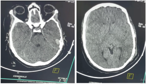
Figure 1: Brain scan showing an extradural hematoma.
The clinical examination on admission found an obnubilated patient with a Glasgow score of 12/15, pupils equal and reactive, presence of clinical stigmata of convulsions (trace of biting of tongues and loss of urine), with a frontal wound of 2 cm sutured, and bilateral palpebral ecchymoses.
In addition, she was hemodynamically stable with a BP: 127/67 on the right and 122/69 on the left, a heart rate of 98 beats per minute with no peripheral signs of shock. On the respiratory level, she was tacypneic at 25 cycles per minute, SpO2 at 99% on room air with normal pleuropulmonary auscultation.
The gynecological examination showed the onset of labor with an open cervix at 3 fingers, and the fetal ultrasound showed a well developed pregnancy, at term with a transverse placenta and an estimated fetal weight of 3800 g, with a fetal heart rate recording without abnormalities.
A brain scan was performed, showing the presence of an extradural hematoma of 0.5 cm with foci of left frontotemporal contusions, the trans-cranial doppler was normal.
The opinion of the neurosurgeons on duty did not retain a surgical indication and the patient was taken to the operating room for fetal extraction.
The patient was placed in supine position with monitoring of blood pressure, heart rate and pulse oxygen saturation
realization of a locoregional anesthesia with bilateral laryngeal block, tracheal block and nebulization of lidocaine 5% and an orotracheal intubation with a fiberscope, with sedation with propofol and curarization, the fetal extraction gave birth to a male newborn in good condition (APGAR score 10/10), with injection of fentanyl after clamping the umbilical cord.
The patient was then transferred to the obstetrical resuscitation service, for prevention and management of secondary cerebral accidents of systemic origin, secondary prevention of convulsions was ensured by levetiracétam.
In front of a normal transcranial doppler, the sedation was stopped 48 hours later and the patient was extubated on the 4th day of her hospitalization after a satisfactory recovery and a respiratory and hemodynamic stability.
Discussion
Traumatic brain injury accounts for 6% to 7% of traumatic injuries in pregnant women. Although less common than abdominal trauma in pregnancy, traumatic brain injuries are more serious in terms of morbidity and mortality. The common causes of trauma were traffic accidents (54.6%), falls (21.8%), violent assaults and burns [1].
Pregnancy should always be suspected in this setting in any woman of childbearing age, unless proven otherwise and confirmed by uterine examination, ultrasound and/or serum bHCG test. Management consists of stabilization of maternal hemodynamics, detailed neurological evaluation, followed by a secondary examination taking into account fetal assessment and the extent of uterine involvement [2].
The best way to treat the fetus is to treat the mother; therefore, the first priority is to stabilize the mother. Initial evaluation includes a thorough assessment, stabilization, and resuscitation to the left lateral decubitus position to prevent hypotensive supine syndrome. The Glasgow Coma Scale (GCS) is recommended. 8 Requires intubation and mechanical ventilation, or both airway control and CASA control [2].
Current recommendations are to maintain normovolemic status with early transfusion of labile blood products rather than crystalloids. If cervical spine trauma is suspected, direct laryngoscopy should be avoided and awake intubation with a fibroscope should be considered. Detailed neurological assessment after maternal hemodynamic stabilization [3].
After initial conditioning, the condition of the fetus and the degree of uterine injury determine subsequent management. Secondary investigations include vaginal and rectal examinations. Gestational age is estimated by uterine height and confirmed by ultrasound. Live fetuses are followed by ERCF. In case of premature labor, tocolysis can be performed. In case of acute fetal distress, a cesarean section is necessary.
Among antiepileptic drugs, monotherapy is preferred, and newer drugs, such as levetiracetam and lamotrigine [4] are now preferred.
The timing of surgery is a huge challenge for neurosurgeons and anesthesiologists. There are conflicting data on the effect of anesthesia in early pregnancy on spontaneous abortion rates, Duncan and colleagues report an increased risk of spontaneous abortion in patients under general anesthesia (hazard ratio 1/4 1.58) [5]. The incidence of spontaneous abortion (15-20%) and congenital anomalies (3-5%) is sufficiently high in the first 13 weeks. Between 13 and 23 weeks of gestation, the uterus is less sensitive to the stimulating effects of surgery and is therefore a safe period for trauma surgery. After 24 weeks of gestation, trauma surgery can result in three complications: supine hypotension, delayed fetal neurodevelopment, and premature birth. Emergency surgery is performed regardless of gestational age.
The prevalence of general anesthesia has decreased significantly and rapid sequence induction should be considered if necessary (deep coma, shock, respiratory distress, etc.) [6].
Conclusion
Pregnancy is a major challenge in trauma care because of the risks to the mother and child and the difficulty in following standard protocols. Management requires a multidisciplinary, conservative, and timely approach to improve maternal and infant outcomes. Postoperative resuscitation should continue with the management of ACSOS and adjust medications accordingly.
References
- Baker BW. Trauma. In: Chestnut DH, ed. Obstetric Anesthesia: Principles and Practice. St Louis: Mosby. 1999: 1041e1050.
- Shah AJ, Kilcline BA. Trauma in pregnancy. Emerg Med Clin North Am. 2003; 21: 615e629.
- Connolly AM, Katz VL, Bash KL, et al. Trauma and pregnancy. Am J Perinatol. 1997; 14: 331e335.
- Flik K, Kloen P, Toro JB, et al. Orthopedic trauma in the pregnant patient. J Am Acad Orthop Surg. 2006; 14: 175e182.
- Reddy SV, Shaik NA, Gualala K. Trauma during pregnancy. J Obstet Anaesth Crit Care. 2012; 2: 3.
- Barker SJ. Anesthesia for trauma. Anesth Analg Suppl. 2003; 96: 1e6.