Case Report
Austin J Clin Case Rep. 2022; 9(3): 1250.
Unilateral Spontaneous Adrenal Haemorrhage in Pregnancy
Seth R and Sinha A*
Department of Radiodiagnosis, Post Graduate Institute of Medical Education & Research, India
*Corresponding author: Anindita Sinha, Additional Professor, Department of Radiodiagnosis, Post Graduate Institute of Medical Education & Research, Chandigarh-160012, India
Received: July 04, 2022; Accepted: August 01, 2022; Published: August 08, 2022
Abstract
Spontaneous Adrenal Haemorrhage (SAH) is a rare condition in which there is haemorrhage in the adrenal gland without any adrenal tumour or history of trauma. It presents as severe flank pain with or without shock depending on the amount of haemorrhage. The initial investigation of choice is ultrasound and the diagnosis can be confirmed by computed tomography or magnetic resonance imaging
We report the case of a pregnant female, presenting with left flank pain and high blood pressure, in the late third trimester. Ultrasonography revealed intra-uterine fetal demise with a large left suprarenal mass. Subsequent imaging by computed tomography and magnetic resonance imaging revealed a heterogeneous left adrenal mass with imaging features suggestive of haemorrhage. The patient delivered spontaneously and her blood pressure returned to normal spontaneously. She was explained about the entity as well as the risks and benefits of surgery for this condition. She did not want to undergo surgery and decided to be on regular follow-up. We repeated the ultrasound every 2 weeks, which showed no increase in the size of the lesion. The patient was asked to continue monthly follow-up for 3 months followed by 3-monthly follow-up by ultrasonography.
This case reiterates the fact that obstetricians should keep a highindex of suspicion for this rare condition after ruling out more common causes of flank pain in pregnancy, and should suspect SAH especially if there is associated severe anemia or shock. The knowledge of this rare entity can help in correct diagnosis and early appropriate treatment.
Keywords: Endocrine (not including DM); Hypertension; Hematologic and Clotting; Preeclampsia/Eclampsia
Case Presentation
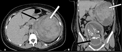
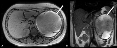
Written and informed consent was obtained. A 25-year-old pregnant lady (primigravida) at 35-weeks-of-gestation presented to emergency with acute left flank pain and vomiting for 2 days. There was no history of fever, bleeding per-vaginum or any urinary complaints. On examination, she was found to have increased blood-pressure (160/100 mm Hg), pulse-rate of 90/min and body-temperature of 37°C with tenderness in the left flank. Her hemogram, renal and liver function tests were within normal limits. However, her serum cortisol was reduced (200 nmol/L). She was managed conservatively with analgesics and antiemetics. Her blood-pressure was controlled by intravenous labetalol. Ultrasonography showed Intra-uterine Fetal Demise (IUFD) with a heterogeneous avascular left suprarenal mass. CT abdomen showed a non-enhancing hyperdense left adrenal mass along with overlapping of foetal calvarial bones (Spalding Sign of IUFD) (Figure 1A & B). MRI findings were also suggestive of adrenal haemorrhage (Figure 2A & B). The patient delivered spontaneously at 37 weeks of gestation and blood-pressure normalized after delivery. A diagnosis of pre-eclampsia and Spontaneous Adrenal Haemorrhage (SAH) was made. The patient refused surgery for the adrenal mass and was discharged on post-partum day 3. She was advised to followup (by ultrasonography) every 2 weeks initially. The follow-up ultrasonographic examinations showed progressive decrease in size of the hematoma suggestive of resolution. There was no recurrence of left flank pain. The patient is still on follow-up and is asymptomatic till date (2 years after SAH).

    Figure 1A & B: A) Contrast enhanced computed-tomography axial image

showing heterogeneous left suprarenal mass (white arrow) with peripheral

calcification (black arrow); B) Coronal reformatted image shows the left

suprarenal mass (white arrow) and the intra-uterine fetus (black arrow).

Overlap of the fetal calvarial bones can also be seen (Spalding sign of

intrauterine fetal demise).

    Figure 2A & B: Axial T1-weighted (A) and T2-weighted (B) magnetic

resonance image showing the left suprarenal mass (white arrows), which

is hyperintense with hypointense central part on both T1 and T2-weighted

images, signifying haemorrhagic contents.
Discussion
SAH refers to haemorrhage occurring in the adrenal gland without any adrenal tumour or trauma. It is an uncommon condition and its incidence varies from 0.14-1.1% (1). However, its exact incidence in pregnancy is not known [2]. Most of the patients present with abdominal pain [1]. If there is extensive bilateral adrenal haemorrhage, symptoms of adrenal insufficiency like vomiting, weakness and diarrhoea may occur [3]. Since it is an uncommon disease with vague clinical presentation, it is difficult to recognise it immediately, which can lead to adrenal crisis and deaths in severe cases. We present a case of SAH in pregnancy who presented with acute flank pain. The imaging findings revealed left sided SAH. The patient was managed conservatively without any recurrence of symptoms (patient is on follow-up and is asymptomatic till date-2 years after SAH).
The exact aetiology of SAH is unclear. Adrenal vein thrombosis and spasm are implicated in some cases [4]. In addition, hypertrophy and hyperplasia of adrenal gland during pregnancy increases its arterial supply, thereby, increasing the chances of SAH during pregnancy [5]. The initial investigation of choice is ultrasonography. On ultrasound, it is seen as a heterogeneous mass with variable echogenicity depending on the stage of hematoma. Colour-doppler confirms that the mass is avascular. MRI is used to confirm the diagnosis and the signal-intensity progresses from hypo to hyperintense (on both T1 and T2-weighted images), with a hypointense-rim in the chronic stage (progression of fresh blood to methaemoglobin and finally hemosiderin) [6].
The differentials of severe flank pain in pregnancy include ovarian torsion, antepartum haemorrhage and abortion. The imaging differentials of adrenal mass include adrenal tumors. However, if MRI shows blood as the only component of the mass, it indicates a benign nature of the mass.
In stable cases, conservative management (correction of coagulopathy and fluid resuscitation) is preferred. Surgery (emergency adrenalectomy with preterm delivery) should be considered if deterioration occurs despite medical management [7]. Arterial embolization may be done as a bridge to surgery, if there is severe haemorrhage [8].
Conclusion
SAH is a rare but serious complication that can occur in pregnancy. It may occur without any history of trauma or adrenal tumour. Ultrasonography is the initial investigation of choice followed by MRI for confirmation. Conservative management is preferred if patient is hemodynamically stable. In cases of pregnant females presenting with flank pain, physicians should keep a high index of suspicion for this condition, after ruling out the more common causes.
Contributor Ship Statement
-Dr Anindita Sinha-planned and conducted the work and submitted the study
-Dr Raghav Seth-Planned and reported the work.
-Dr Anindita Sinha is the overall guarantor.
Funding
This study was not supported by any funding.
Conflict of Interest
The authors declare that they have no conflict of interest.
Consent for Publication
Consent for publication was obtained for every individual person’s data included in the study.
References
- Imga NN, Tutuncu Y, Tuna MM, Dogan BA, Berker D, Guler S. Idiopathic Spontaneous Adrenal Hemorrhage in the Third Trimester of Pregnancy. Case Reports in Medicine. 2013; 2013: 1-2.
- Gavrilova-Jordan L, Edmister WB, Farrell MA, Watson WJ. Spontaneous Adrenal Hemorrhage During Pregnancy: A Review of the Literature and a Case Report of Successful Conservative Management. Obstetrical & Gynecological Survey. 2005; 60: 191-195.
- Wani MS, Naikoo ZA, Malik MA, Bhat AH, Wani MA, Qadri SA. Spontaneous adrenal hemorrhage during pregnancy: review of literature and case report of successful conservative management. Journal of the Turkish German Gynecological Association. 2011; 12: 263-265.
- Kovacs KA, Lam YM, Pater JL. Bilateral massive adrenal hemorrhage. Assessment of putative risk factors by the case-control method. Medicine (Baltimore). 2001; 80: 45–53.
- Kadhem S, Ebrahem R, Munguti C, Mortada R. Spontaneous Unilateral Adrenal Hemorrhage in Pregnancy. Cureus. 2017; 9.
- Kawashima A, Sandler CM, Ernst RD, Takahashi N, Roubidoux MA, Goldman SM, et al. Imaging of nontraumatic hemorrhage of the adrenal gland. Radiographics : a review publication of the Radiological Society of North America, Inc. 1999; 19: 949-963.
- Bockorny B, Posteraro A, Bilgrami S. Bilateral spontaneous adrenal hemorrhage during pregnancy. Obstetrics and gynecology. 2012; 120: 377- 381.
- Patel A, Downing R, Vijay S. Spontaneous Rupture of the Adrenal Artery Successfully Treated Using the Endovascular Approach. Vascular and Endovascular Surgery. 2013; 47: 124-127.