Case Report
Austin J Clin Case Rep. 2022; 9(3): 1251.
Thorn in my Side - A Case of Entrapped, Intra-uterine Foreign Body and Delayed, Atypical Pelvic Inflammatory Disease
Aboda A*, Kim H, Xu B and McCully B
Department of Obstetrics & Gynaecology, Mildura Base Public Hospital, Australia
*Corresponding author: Ayman Aboda, Department of Obstetrics & Gynaecology at Mildura Base Public Hospital, Mildura VIC 3500, Australia
Received: July 06, 2022; Accepted: August 05, 2022; Published: August 12, 2022
Abstract
Uterine foreign bodies are a rare, but important cause of infertility and pelvic pain. Due to their relative scarcity, the diagnosis may be either missed or simply not considered as contributory to aetiology when patients present with symptoms. We report the case of a 42-year-old woman diagnosed with Atypical Pelvic Inflammatory Disease (APID). She presented with lower abdominal pain, vaginal discharge and inflammatory markers consistent with acute infection. Paradoxically, diagnostic imaging revealed an unsuspected intrauterine foreign body. The significance of this to the presenting complaint was initially uncertain however conservative management with antibiotics alone was sufficient to allow complete recovery. Later, the patient consented to operative hysteroscopy which identified and removed the object which was shown by histology to be a residual bone fragment derived most probably, from retained products of conception. The patient was noted to have had a surgical termination of pregnancy 5 years prior.
Keywords: Termination of pregnancy; Foreign body; Intra-uterine; Bony fragment; Atypical pelvic inflammatory disease; Secondary infertility; Contraception
Case Presentation
A 42-year-old woman attended the emergency department with a two-week history of right-sided pelvic pain, dyspareunia, dysuria, and leukorrhea. She reported having multiple sexual partners, a history of IV drug use, and previous chlamydia infection treated one year prior. Her obstetric history included three vaginal deliveries and two surgical terminations of pregnancy, the most recent performed 5 years earlier at 17 weeks gestation. Her menstrual cycle was regular with normal bleeding associated with occasional mid cycle spotting and pelvic discomfort with coitus. Her cervical screening was normal and up to date.
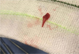
On examination, she was a febrile with normal vital signs. There was mild abdominal tenderness with palpation but no signs of peritonism. Pelvic examination revealed copious amounts of white, non- offensive discharge. The cervix and external os were normal with mild pelvic discomfort elicited by cervical motion. Blood results were consistent with an acute inflammatory process showing increased white cells (WCC) 14.2 x10^9/L [Normal range 4.0-12.0], neutrophilia, 9.1x10^9/L [NR: 2.0-8.0], and elevated C Reactive Protein (CRP) 64.2mg/L [NR: <5.0]. Her urine culture showed no growth. Swabs for Gonorrhoea and Chlamydia were negative however anaerobes consistent with bacterial vaginosis were identified on high vaginal swab. Curiously, an abdominal CT scan revealed an unsuspected foreign body in the lower uterine cavity. It did not have the characteristics of an Intra-Uterine Contraceptive Device (IUCD) and was found with subsequent transvaginal pelvic ultrasound to be approximately 11mm in size. The reporting sonographer was not able to ascertain its origin. The patient was questioned in more detail but remained certain that she had never used an IUCD and had not engaged in any form of unusual sexual play. She was diagnosed with Atypical PID and was managed expectantly with outpatient antibiotic treatment for two-weeks. At the time of review, the suspicion of an intra- uterine foreign body was again discussed. The patient described her past previous TOP but could not remember if any complications occurred afterwards. She consented to surgical Examination Under Anaesthesia (EUA) including hysteroscopy with curettage to locate and remove the object. The procedure was performed without complication. A fragment of white, osseous material was retrieved and later identified as bone with stromal smooth muscle by histopathology, (Figure 1). At the patient’s request, Mirena was placed at the time of the procedure.

Figure 1: Foreign Body.
Discussion
Pelvic Inflammatory Disease (PID) is one of the oldest infections known to women. Despite primary health interventions and community education, it remains today a common concern. Patients can present with a spectrum of clinical signs and symptoms [1]. They may do so acutely with fulminating pain and sepsis associated with malodorous vaginal discharge; or, as with the example of our patient, more indolent symptoms that may arise many weeks or months after a possible exposing event [2-4]. These latter, atypical presentations are significant for without the provocation for immediate care, delayed diagnosis and treatment may allow the progress of longterm sequalae such as pelvic and tubal inflammatory disease leading to chronic pain, infertility and risks of ectopic pregnancy [5]. Early suspicion of atypical PID is therefore vital if we are to help reduce the burden that this disease can wreak in women of reproductive age.
As much as 85% of PID is caused by sexually transmitted pathogens. Remaining cases are often associated with enteric organisms [3]. When cultures from high or low vaginal swabs are negative, other less common causes may include the sequalae of adnexal pathology or, as in our case, the effects, though invariably shrouded, of an intra-uterine foreign body. Such instances occur rarely, and are most often associated with initial placement or prolonged retention of an Intrauterine Contraceptive Device (IUCD) [6-8]. Our case is unusual, in that it reports an altogether unexpected mechanism of foreign body retention. We know from histology, that the material was bony and was most likely of foetal origin arising from remnants retained from a previous failed pregnancy. Reports from the literature often cite spontaneous miscarriage or iatrogenic termination as the reason for such earlier loss. In our report, the patient confirmed a surgical termination 5 years earlier. The procedure had been performed at 17 weeks and though uncomplicated, may well have led to retained products of conception that remained quiescent or at least, asymptomatic until the current presentation. Anomalous, intra-uterine bony material may also occur de novo as a rare form of endometrial metaplasia [9-11]. In such examples, it can be distinguished from foetal remnants by its continuity with underlying endometrial stroma [12]. This was not the case in our report, thus affirming the likely sodality of our findings to the patient’s previous obstetric history.
Why is this important? We note that our patient had Implanon inserted following her termination. This was removed 2 years later, following which no specific precautions were taken to reduce pregnancy risk. The patient did not conceive again although the opportunity to do so was certainly present. She was not however upset by this, nor aware of any failure that it might imply. We know however, that for many women, secondary infertility is a leading presentation associated with prolonged sequestration of intrauterine foreign bodies. Once removed, restoration to normal fertility follows soon afterwards. Whilst this was not a concern for our patient, we propose that for as long as the condition persists undiagnosed, there will be some grievance to normal fertility leading to unexplained delay or interruption of normal childbearing and ultimately, a cumulative risk of age-related disease that could otherwise been mitigated if earlier diagnosis had occurred. Additionally, while the material remains undiscovered, patients are at risk of chronic pelvic pathology which may itself inhibit or complicate future fecundity. As we have seen, symptoms may be muted. Patients tend to present with secondary infertility rather than acute infective pathology [13-17] and for this reason, may elude medical care for many years beyond the exposure event [18]. As in our case, definitive management requires surgical removal of the foreign body [19-22] to restore normal endometrial implantation potential and inflammatory risk profile. Having done so, return to normal fertility is an immediate and for many, a much sought-after boon of successful care.
As we prepared this report, we considered the history of deliberately placed, intra-uterine bodies as agents of desired fertility control. We wondered if inspiration for the innovation of such devices may have been spurred by the observation of cases such as ours, where the discovery of a retained object was later recognized as causative, or contributory to, fertility impairment. It is an eloquent premise, but there seems little to support such catalyst for innovation. We note however that in 400 B.C., Hippocrates, the father of medicine, was said to have inserted small, inert pessaries into the uterus of fertile women to help protect them from unwanted pregnancy [23,24]. Interestingly, he also suggested the use of copper tainted water to induce prolonged infertility, an observation reiterated nearly 2,500 years later when researchers found that copper inside the womb of rabbits would lead to infertility [25]. More apposite with our discussion, nomadic Bedouin traders were famed for their habit of putting small stones into the wombs of female camels to keep them sterile for the duration of long treks [26]. Once removed, the camels returned easily to their normal breeding patterns. The first successful intra-uterine device for human contraception was developed by German physician, Dr. Richter in Germany in 1909 [27]. We may never know his inspirations for doing so, but they seem unlikely to have arisen through any curiosities of uterine misadventure. Indeed, his device was sited at the opening of the cervix presumably to impede sperm passage rather than castigate the endometrial environment. Never-the-less, it seeded innovation and with numerous iterations since, it has led to the intra-uterine devices we have today which provide safe, reliable choice for reproductive wellbeing.
Conclusion
Intrauterine foreign bodies are most commonly iatrogenic. More rarely, they can present as remnants residual of previous pregnancy loss. Significantly, the material may linger undiscovered for many months or even years without causing identifiable symptoms and yet, may still be associated with progressive risk of chronic pelvic disease and infertility. The suspicion of a retained foreign body should therefore be included as part of any diagnostic work up for patients presenting with symptoms of pelvic infection or unexpected fertility failure, particularly in the setting of prior pregnancy loss. We present this case to heighten such awareness.
References
- LK Jennings, DM Krywko Pelvic inflammatory disease. Stat Pearls [Internet]. 2022.
- Al-kuran O, Al-Mehaisen L, Alduraidi H, Al-Husban N, Attarakih B, Sultan A, et al. How prevalent are symptoms and risk factors of pelvic inflammatory disease in a sexually conservative population. Reproductive Health. 2021; 18.
- Brunham RC, Gottlieb SL, Paavonen J. Pelvic inflammatory disease. The New England journal of medicine. 2015; 372: 2039-2048.
- Curry A, Williams T, Penny ML. Pelvic Inflammatory Disease: Diagnosis, Management, and Prevention. American family physician. 2019; 100: 357- 364.
- Ravel J, Moreno I, SimÓn C. Bacterial Vaginosis and Its Association with Infertility, Endometritis, and Pelvic Inflammatory Disease. American journal of obstetrics and gynecology. 2020; 224: 251-257.
- K Ketvertis, Intrauterine Device EL Lanzola. Stat Pearls [Internet]. 2022.
- Birgisson NE, Zhao Q, Secura GM, Madden T, Peipert JF. Positive Testing for Neisseria gonorrhoeae and Chlamydia trachomatis and the Risk of Pelvic Inflammatory Disease in IUD Users. Journal of women’s health. 2015; 24: 354-359.
- Levin G, Dior UP, Gilad R, Benshushan A, Shushan A, Rottenstreich A. Pelvic inflammatory disease among users and non-users of an intrauterine device. Journal of Obstetrics and Gynaecology. 2020; 41: 118-123.
- B Abdull Gaffar, A AlMulla. Endometrial calcifications. International Journal of Surgical. 2020.
- Peart J, Pillay S. Endometrial osseous metaplasia in a patient with secondary infertility referred for hysterosalpingogram with ultrasound. Journal of Medical Imaging and Radiation Oncology. 2021; 65: 909-910.
- Garzon S, Laganà AS, Carugno J, Font EC, Jimenez J, Kar S, et al. Osseous metaplasia of the endometrium: A multicenter retrospective study. European journal of obstetrics, gynecology, and reproductive biology. 2021; 265: 150- 155.
- Patil S, Narchal S, Paricharak D, More S. Endometrial Osseous Metaplasia: Case Report with Literature Review. Annals of Medical and Health Sciences Research. 2013; 3: 10.
- Cao J, Grubb C, Khurshid M, Gumma A. Retained fetal bone post abortion causing infertility. Obstetrics and Gynaecology. 2021.
- SR Sharma, Retained Foetal Bone Fragment Masquerading as Foreign Body Causing Secondary Infertility SM Toshniwal. ABS Tembhare. 2022.
- JA Lawrence, U Maskey, A Agolli, Charmy Parikh. Secondary infertility due to fetal bone retention: A systematic literature review. Sultan Qaboos University Medical Journal [SQUM]. 2022.
- Izhar R, Husain S, Tahir S, Husain S. Secondary Infertility due to Retained Fetal Bones Diagnosed via Saline Sonography. Journal of the College of Physicians and Surgeons--Pakistan: JCPSP. 2016; 26: 861-862.
- IO Awowole, OO Badejoko, EO Ayegbusi, OO Allen and OM Loto. Infertility Due to Prolonged Retention of Fetal Bones: A Case Series. Journal of Gynecologic Surgery. 2022.
- Al-Shawaf T, Brown J, Keegan C. Retention of fetal bones 8 years following termination of pregnancy. Ultrasound in Obstetrics and Gynecology. 1992; 2: 61-63.
- Amodeo S, Iannone V, Borriello M, Giambanco L. Hysteroscopic management of endometrial osseous metaplasia mimicking a foreign body. Journal of minimally invasive gynecology. 2021; 28: 1673-1674.
- J Arun, N Chavali, S Bechler, D Klindt, A.L Vikins. Hysteroscopic Removal of Foreign Body. Journal of Minimally. 2021.
- VV Mishra, SB Solanki, R Aggarwal, M Anusha. Intrauterine Retained Bone Fragments Causing Secondary Infertility: A Case Report. J Med Case Rep. 2022; 3.
- Catinon M, Roux E, Auroux A, Trunfio-Sfarghiu A, Lauro-Colleaux C, Watkin E, et al. Confirmation of the systematic presence of tin particles in fallopian tubes or uterine horns of Essure implant explanted patients: A study of 18 cases with the same pathological process. Journal of trace elements in medicine and biology: organ of the Society for Minerals and Trace Elements. 2021; 69: 126891.
- Intrauterine devices. Fam Plann Inf Serv. 1978; 1: 1-50.
- Bujalkova M. Birth control in antiquity. Bratislavske lekarske listy. 2007;108(3):163-6.
- Zipper JA, Tatum HJ, Pastene L, Medel M, Rivera M. Metallic copper as an intrauterine contraceptice adjunct to the “T” device. American journal of obstetrics and gynecology. 1969; 105: 1274-1278.
- Wessel GM. Of camels, silkworms, and contraception. Molecular Reproduction and Development. 2014; 81: Fmi-Fmi.
- Richter R. Ein Mittel zur Verhütung der Konzeption. Deutsche Medizinische Wochenschrift. 1909; 35: 1525-1527.