Clinical Image
Austin J Clin Case Rep. 2022; 9(4): 1253.
Gastric Diverticula: An Uncommon Finding
Adil H*, Choayb S, El Fenni J and En-Nafaa I
Department of Radiology, Mohammed V Military Teaching Hospital, Faculty of Medicine and Pharmacy, Mohammed V University, Rabat, Morocco
*Corresponding author: Adil Hajar, Department of radiology, Mohammed V Military Teaching Hospital, Faculty of Medicine and Pharmacy, Mohammed V University, Rabat, Morocco
Received: July 29, 2022; Accepted: August 19, 2022; Published: August 26, 2022
Clinical Image
Gastric Diverticula (GD) are extremely uncommon. They define as an out pouching of the gastric wall to form a sac-like structure. They can be congenital or acquired and are mostly located on the posterior surface of the gastric fundus.
GD is asymptomatic in general; however, they may present with discomfort or vague abdominal pain. Rarely, GD can be revealed by a complication, such as ulceration, perforation, or hemorrhage.
Abdominal CT scan demonstrates a thin-walled, cystic lesion located in the posterior wall of the fundic region. It may sometimes contain air bubbles or air-fluid level.
Communication with the gastric lumen is easily demonstrated by ingesting a water-soluble contrast.
Differential diagnosis includes adrenal, pancreatic, and renal cysts, duplication cysts, and bowel diverticula.

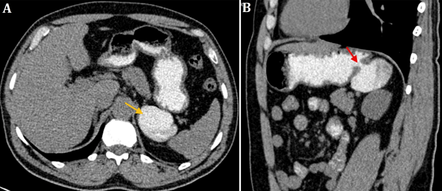
    Figure 1: Axial (A) and sagittal reformatted (B) images demonstrating a

gastric diverticulum arising from the posterior surface of the fundus, and

filled with the oral contrast agent (yellow arrow). The red yellow indicates the

communication between the diverticulum and the gastric lumen.